

ADVERTISEMENT
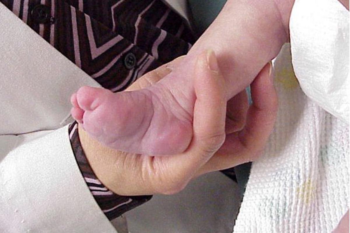
Current Concepts In Clubfoot Treatment
Given the challenges of treating talipes equinovarus, this author offers a closer look at the evolution of the Ponseti method, keys to successful treatment, pertinent factors that contribute to adverse results and proactive steps to reduce complication risk.
 Talipes equinovarus remains a clinically difficult problem because of the complexity of the morbid anatomy and narrow treatment options that are limited to manipulation with minor surgery at one end and extensive surgical releases at the other end. The goals and objectives of management of this deformity are a supple, painless, high functional and plantigrade foot with enough motion in the ankle and rearfoot to achieve an efficient heel to toe gait pattern, and permit wearing acceptable shoe styles. Physicians have refined both surgical and nonsurgical techniques over the years in an attempt to try to meet these objectives.1
Disenchantment with extensive surgical management of clubfoot reached its peak in the early 1990s.2 Undesirable outcomes from surgical release included severe overcorrection, major joint surface injury, avascular necrosis of the tarsal structures, subluxed weightbearing articulations, soft tissue contractures and problems with extensive surgical scars.3
Nonoperative techniques have evolved throughout the last century. Beginning with Hoke and then Kite, surgeons made attempts to correct the deformity through serial casting.2 Kite’s technique was based on isolation of the three major components of the clubfoot. The first component is adduction of the forefoot (thought by Kite to be similar to metatarsus adductus). The second component is inversion of the subtalar joint complex. The third component is equinus at the ankle.
Kite attempted to correct these one at a time and independently beginning with abducting the forefoot, subsequent everting of the rearfoot complex and finally management of equinus. This was only partly successful and many of the patients remained incompletely or totally unreduced. The infants wore casting until they were about 9 months of age and then underwent increasingly complex major releases of the residual deformity.
 We can trace the reason why the technique was less than successful back to isolating each of the components of the deformity and attempting to treat them independently. As more knowledge of rearfoot biomechanics emerged, it became clear that it is not possible to isolate the rearfoot varus from the forefoot adductus since one must manage these together and consider the entire talocalcaneonavicular joint complex (the acetabulum pedis) as a unit.
In short, we can trace the less than successful results with Kite’s method back to a failure to fully appreciate the pathological anatomy of the clubfoot deformity. We now recognize that the overall equinus position of the foot is the sum of the ankle equinus plus the plantarflexion within the foot as the result of talocalcaneonavicular supination.
How Ponseti Contributed To The Understanding Of Clubfoot
Ponseti had a much better understanding of the pathological anatomy. He recognized that the forefoot abnormality was actually an adductovalgus deformity. Attempts to move the forefoot in isolation in the transverse plane would not only fail to reduce the deformity but actually accentuate a medial column cavus deformity. He also recognized that the apparently inverted position of the rearfoot was really a pathological supination of the entire talocalcaneonavicular joint complex.
 In order to reduce the deformity, it is necessary to manipulate the entire complex to bring the navicular up and lateral to its more anatomical position on the talar head, move the distal calcaneus from a medial and plantar position to a more anatomical dorsal and lateral location along the lateral talar neck, and also allow the calcaneus to translate in the lateral direction around a more or less vertical axis.
Why is the Achilles tenotomy necessary and sectioning of ligaments is not? Based on the physiology of the soft tissues, Ponseti realized that ligament and capsule can gradually stretch with persistent but gentle manipulation and holding. Tendon cannot stretch. It was his contention that one should address unyielding equinus by tenotomy of the tendo-Achilles just above its insertion into the calcaneus. The number of casts required and higher Pirani and Dimeglio scores predict the need for Achilles tenotomy.4 Based on personal experience as well as the current literature, tenotomy is indicated in at least 85 to 97 percent of cases.5,6 Possible complications from the tendo-Achilles tenotomy include trauma to the peroneal and posterior tibial vasculature, damage to the sural nerve and lesser saphenous vein, and incomplete release of the tendon.5,7
Performing an in-office tenotomy without patient sedation or general anesthesia is safe, but performing the procedure in the operating room with the patient under general anesthesia makes successful application of the postoperative cast easier than attempting application in an uncooperative infant.8 Since the need for tenotomy is so great in this population, we cannot consider the Ponseti technique to be totally non-surgical. Since the acceptance of the method, radical surgery is now confined to resistant cases.
By avoiding major surgical invasion of the posterior ankle compartment, physicians can successfully manage equinus deformity without producing large surgical scars and deep tissue fibrosis. Since the 1990s, Ponseti’s technique is internationally regarded as the best management of talipes equinovarus.9-12 Advantages of the technique include better range of motion, less pain, less need for extensive surgery and less joint deformity.13,14
 Manipulation achieves the initial reduction of the deformity but in order to maintain the reduction, long-term bracing is necessary. The purpose of bracing is to prevent recurrence or relapse by retaining the pronation of the talocalcaneonavicular complex through abduction or external rotation of the foot. However, this type of bracing does little to preserve ankle motion in the sagittal plane. Ankle-foot orthoses (AFO) may maintain ankle position but do not maintain the midfoot correction. Recurrence of equinus is best regarded as sagittal plane deformity.
Where The Ponseti Method Is Limited
In spite of the greater success of Ponseti’s technique, there are still therapeutic failures. There are a number of explanations for this.
First, it is essential to accept that talipes equinovarus is an anatomical lesion and not a diagnosis. It is possible to classify talipes equinovarus a number of different ways based on etiology and the morbid anatomy.
 Thompson and Simons recognized that there are a number of forms of talipes equinovarus.15 The idiopathic form comprises the largest number of the cases. However, they recognized that there are several other types that we need to acknowledge and classify. Postural and positional talipes equinovarus theoretically develops late in gestational time and results from either packing abnormalities (therefore, not a “parts” problem) and possibly in some cases secondary to muscle imbalance. The teratologic or teratogenic form occurs in association with other types of orthopedic abnormalities such as arthrogryposis, myelomeningocele and tethered cord. The syndromic form is associated with multisystem pathologies such as myotonic dystrophy and other named syndromes.
Ponseti divided talipes equinovarus into two broad types (see “A Quick Guide To Clubfoot Classification For The Ponseti Technique” below at right). Typical talipes equinovarus corresponded to variations of idiopathic talipes equinovarus. These include positional deformities, infants whose treatment was delayed over six months, recurrence of idiopathic clubfoot and clubfoot treated unsuccessfully with other techniques.
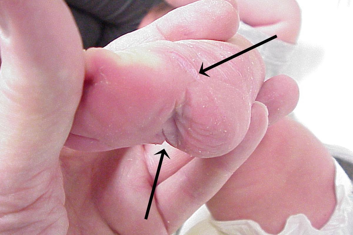
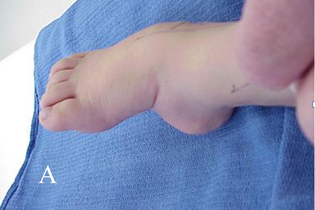
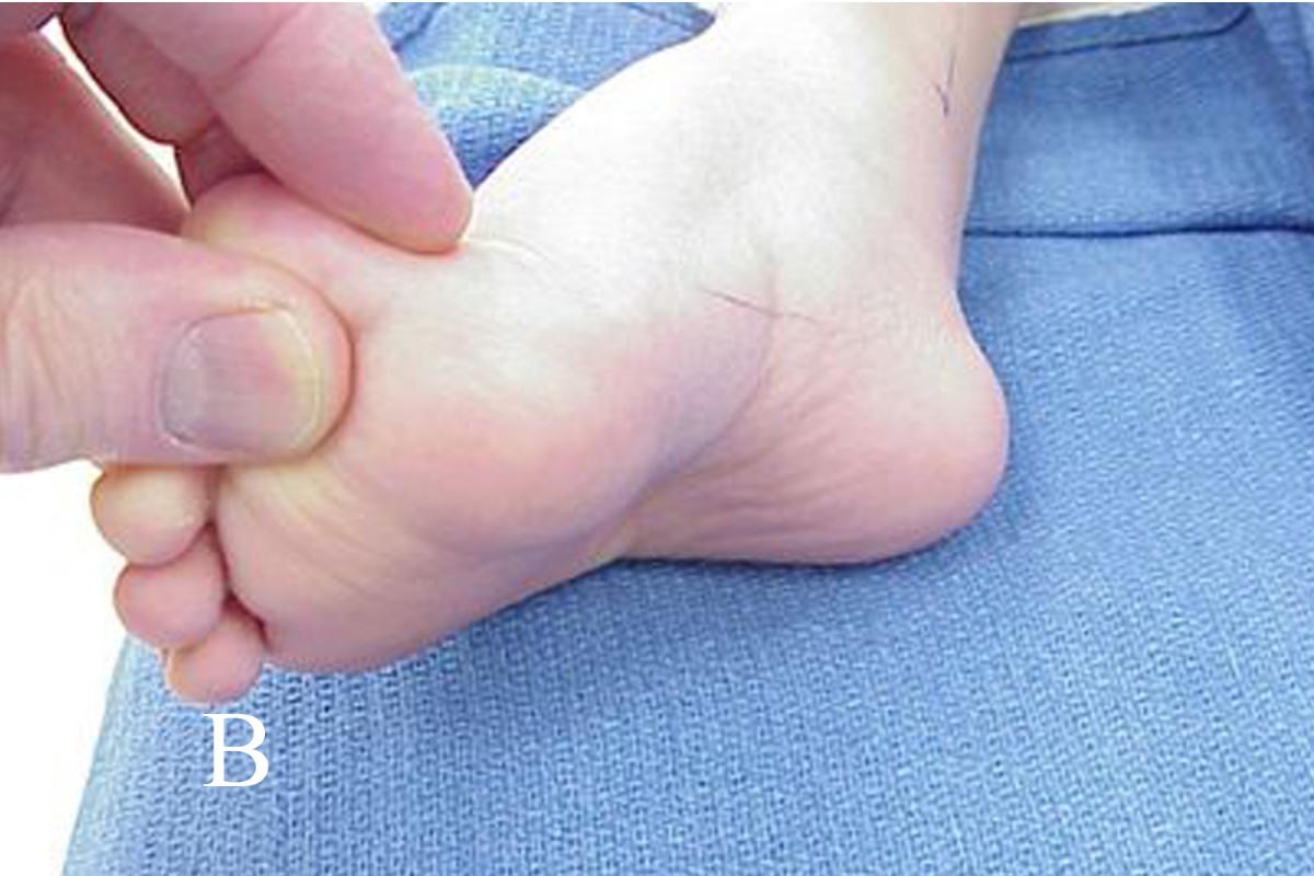
 Ponseti also recognized an atypical form of talipes equinovarus. The major features of atypical talipes equinovarus are plantarflexion of both medial and lateral columns, short foot, rigid equinus, a deep plantar and medial midfoot crease, and a deep crease above the posterior heel. He considered these to be resistant, some syndromic, others teratologic, some neurogenic and occasionally iatrogenic. He modified the manipulation and casting protocol for these feet.
Mater and colleagues studied 11 atypical clubfeet.16 The authors noted these feet required more casts, were more likely to need an Achilles tenotomy and had a higher risk of relapse (53 percent), resulting in a higher incidence of major surgery. Gurnett and coworkers reached similar conclusions.17 Residual equinus after Achilles tenotomy is highly suggestive of a need for additional surgery in the future.18
 What Factors Influence The Prognosis For Success?
The child’s prognosis first depends on the type of clubfoot deformity and the classification. Fortunately, both classifications group most talipes equinovarus as idiopathic. The presumption is that there is no underlying etiology or comorbidity that might adversely affect prognosis. The exact cause of idiopathic talipes equinovarus remains open to debate and there is probably no single etiologic factor.
Of great interest, there has been progress in an attempt to identify a gene or a series of genes that may predispose children to talipes equinovarus. That would strengthen the position of talipes equinovarus as a genetically mediated, hereditary congenital deformity.19-23
One can use the Ponseti technique to successfully manage the other forms of talipes equinovarus. Teratological forms include myelomeningocele, tethered cord and arthrogryposis. Mater and colleagues evaluated the Ponseti technique for management of talipes equinovarus in patients with myelomeningocele.24 Authors concluded that it was an effective first-line therapy but noted that more casts were required, and there was a higher incidence of relapse. Jackson and coworkers studied the Ponseti technique in patients with tethered cord clubfoot and concluded that the method is valid, but the patients required more casts and had a 42 percent recurrence rate.10 Several studies concluded that the Ponseti technique is a good first-line treatment for arthrogrypotic clubfoot.25-27 The Ponseti technique is also effective after unsuccessful posteromedial release.28,29
Second, although seemingly simplistic and easy, the Ponseti method requires training in order to perform the technique successfully. Strict adherence to the method’s principles is absolutely essential.30 Surgeons who wish to pursue this form of therapy should obtain training from a specialist who is knowledgeable, has experience and is skilled in the technique.
How To Perform The Ponseti Technique Successfully
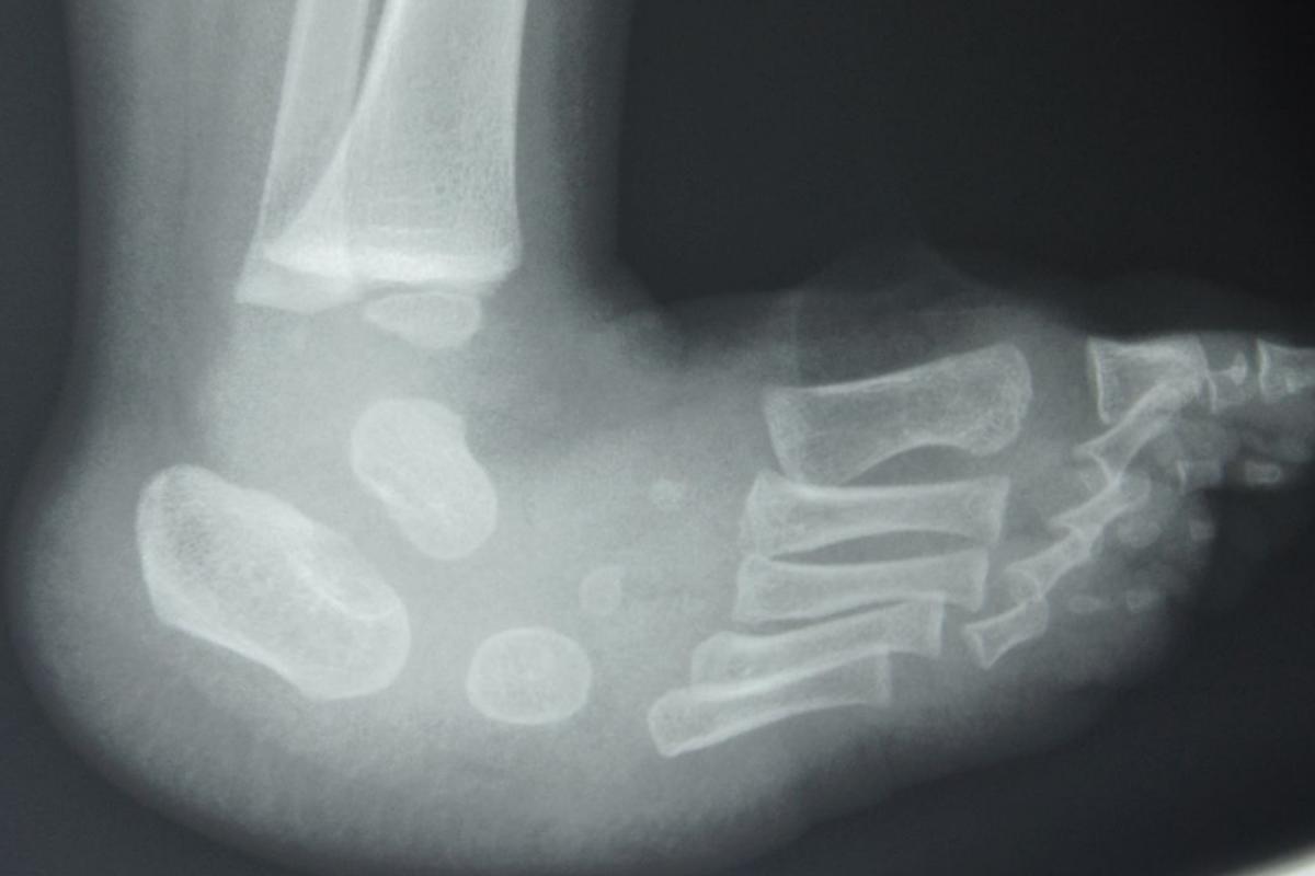
Briefly, treatment begins with identifying the morbid anatomy. Radiographs are unreliable and unlikely to enter into initial decision-making so they are rarely necessary to begin treatment. The surgeon initially palpates the ankle area at the malleoli and then brings his or her fingers and thumb distally to identify the talar head, noting that the navicular is displaced on the plantar and medial aspect of it.
 After establishing landmarks, the next step is to reduce the cavus deformity of the medial column by supinating the forefoot. When this occurs, the next step is to bring the navicular as well as the remaining tarsal structures back into anatomical position by moving the navicular from its inferior and medial position on the talar head upward and lateral to its correct anatomical position on the head of the talus. When this occurs, the distal portion of the calcaneus moves from its position under the talar head and comes laterally and upward while at the same time translating the body of the calcaneus about a vertical axis so the posterior portion of the calcaneus everts and moves medially.
Following this, direct attention to the equinus component.27 My personal experience has been that most of our patients do not respond completely to attempts to cast the foot into a dorsiflexed position. Continued efforts will either breach the foot through the midfoot articulations, damage the talar dome or both. With very few exceptions, these patients require a heel cord tenotomy.
What Are Some Of The Possible Adverse Results?
 Overcorrection of the foot using the Ponseti technique is an extremely rare event. Very little information is available in the literature. In one single series, Hayes and colleagues reported a 12 percent overcorrection with this technique.13 The majority of problems are incompletely corrected deformities, recurrences or relapses. Hosseinzadeh and coworkers noted that researchers have reported a 26 to 42 percent recurrence rate, identifying this as a substantial problem.32 Recurrence of equinus and dynamic forefoot supination are often the first signs of relapse.33
There are a number of factors triggering these adverse results. First, we can trace many of the complications back to an incorrect understanding of the anatomical deformity. Less experienced surgeons tend to take components from the Kite technique and incorporate them into the Ponseti method. This results in a less than desirable outcome because the anatomical concept of Kite’s technique was faulty from its inception. The best way to avoid this potential complication is to adhere strictly to the Ponseti technique.
The second reason for failure to maintain full correction is in the casting technique itself. In order to maintain transverse positioning of the rearfoot, it is necessary to have an above-the-knee cast with the knee flexed a sufficient amount to keep the foot externally rotated. Short leg casts simply do not work. Maripuri and colleagues undertook a randomized trial to evaluate above-the-knee casts in comparison to below-the-knee casts.34 The authors ended the study early for ethical reasons because of the failure rate in the below-the-knee cast group. If the cast does not extend above the flexed knee, maintenance of the manipulation cannot occur.
Third, allowing the foot to simply pronate without maintaining a better forefoot position results in an increase in forefoot cavus and the calcaneus does not evert and abduct. This casting error results in external rotation of the foot in the ankle mortise instead of abducting and everting the calcaneus. One should abduct the foot in some degree of supination and flexion with counter pressure over the lateral talar head.
Fourth, until one has correctly aligned the forefoot and rearfoot, it is inappropriate to treat ankle equinus. Doing so has several undesirable consequences. Casting an unyielding equinus deformity will eventually damage the talar trochlear surface, making it flat and causing it to lose its sphericity. Ankle dorsiflexion will be mechanically limited. Rocker bottom deformity results from attempting to dorsiflex the rearfoot prematurely against the unyielding tendo-Achilles. Inflexible equinus causes the midfoot to dorsiflex against the rearfoot along the lateral column while the calcaneus remains in a plantarflexed position. This breaches the foot through the calcaneocuboid joint as well as the fourth and fifth metatarsal cuboid articulations.
Fifth, there is one other problem in addressing the equinus by tenotomy before the rearfoot is fully corrected. By doing so, the surgeon loses the foot stability provided by the equinus that is necessary to maintain the corrected position in the cast. Before the equinus is correctable, the anterior process of the calcaneus has to abduct from its position under the talus and the foot abduction position in the cast has to be on the order of about 60 degrees. Ideally, the calcaneus should be in slight valgus but a neutral calcaneus is acceptable.
 Sixth, tenotomy errors include performing the tenotomy before correcting the heel and forefoot, and potentially failure to complete the tenotomy. Some studies suggest that incomplete sectioning of the tendo-Achilles may still yield a successful result.35 A new issue that is beginning to emerge among surgeons is that some either do not believe in or are reluctant to perform the tenotomy. Additionally, the surgeon may erroneously believe that the equinus is corrected when it is not.
Seventh, pressure sores may develop as the result of poor casting technique. One has to apply the casts very gently and very carefully in order to prevent this complication. Other potential complications of casting include moisture lesions, hematoma and dermatitis.36 After the tenotomy, the cast may conceal unsuspected bleeding.
Eighth, another reason for failure of the technique may have to do with allowing the parents to take the cast off at home. My personal bias is to remove the cast in the office. Otherwise, the surgeon is never really sure when the cast came off. It may have come off an hour or two before the appointment as instructed, the day before or perhaps even the day the cast went on. This relinquishes an important therapeutic control to the parents that they should not have.
Ninth, it is almost universally agreed that the greatest cause both for early failure of the Ponseti technique and the likelihood of recurrence is failure to appropriately brace after successful cast reduction.37-44 The protocol calls for full-time bracing for three months after the reduction followed by nighttime bracing for three or four years afterward. Strict adherence with bracing does not guarantee that there will be no relapse. Some totally adherent infants recur while some with less than full adherence do not.40 Non-adherence with the foot abduction orthosis protocol may result in a 7.9 times higher need for additional major surgery.45
In summary, the risks for recurrence include failure to comply with bracing, parental educational level of high school or less, increase in the number of casts required for reduction and failure to follow patients up at appropriate intervals.46,47 Other issues for brace non-adherence include positioning the shoes on the bar, infant comfort, affordability and delay in modifying the brace as the infant grows. The costs in parental time for examinations and regular follow up need to be factored in as well.48
How To Handle Relapse
It is hard to determine whether failure of complete reduction is due to recurrence or accepting incomplete reduction at the end of the initial treatment. It is necessary to follow up regularly to diagnose early true recurrence and resume casting to reestablish correction.
True relapse is characterized by any degree of loss of rearfoot abduction, loss of ankle dorsiflexion and rigid subtalar varus.49 One must identify these early to reestablish casting. Once true relapse has occurred, it is necessary to determine whether relapse is secondary to muscle imbalance or some other manageable cause. If relapse is secondary to muscle imbalance, the treatment is repeat reduction in casts followed by transfer of the tibialis anterior to the level of the third cuneiform and possibly repeat Achilles lengthening. On occasion, plantar release and fractional lengthening of the tibialis posterior may be indicated. With additional complex deformity, management becomes “à la carte.” It is important to resume casting and bracing the same way as the patient had in infancy. Tibialis anterior transfer may not prevent additional recurrence in known or unsuspected neuromuscular disease. Lovell and Morcuende reported four cases of late clubfoot relapse associated with neuromuscular disease (myotonic dystrophy, multicore disease, Charcot-Marie-Tooth 1A and myasthenia gravis).50
Taking Steps To Reduce Complication Risk
In order to minimize undercorrection and other potential complications including recurrence, it is important for the surgeon to be well trained in the Ponseti technique. It is also important to adhere strictly to the principles and resist the urge to embellish or change the technique.51 Authors have proposed accelerated casting by decreasing the interval between cast changes to several times a week and the result is a decrease in the time required to achieve reduction.52-54 This is not a change in the technique. It simply represents a refinement in the process. Adding procedures specific for individual cases is also acceptable as long as physicians observe the basics.
 Other changes are alterations in the basic principles. Ponseti advocated plaster of Paris because of its superior molding. Other forms of hard or softer cast materials are available, and multiple authors have discussed the use of these materials in the literature.38,55
One study evaluated the results of very early transfer of tibialis anterior tendon and found that bracing time was reduced, but possible weakening of dorsiflexion could be a potential issue.56 Grigoriou and colleagues studied performing a posterior capsulotomy and open lengthening of the tendo-Achilles, and found that these procedures did not improve dorsiflexion of the ankle or alter the recurrence rate.57 Their recommendation was to confine this surgery to tenotomy alone. Kumar and Gopalakrishina undertook a study to modify the Ponseti technique by performing the Achilles tenotomy before the application of the first cast.58 Persistently high Dimeglio or Pirani scores immediately after tenotomy were predictors of failure.
Other Pertinent Considerations
The protocol has always been to initiate treatment as soon after birth as possible. Problems with very early treatment include cast slipping, skin issues from casting, skin issues with bracing, early non-adherence with bracing and early initial relapse. We should no longer regard treatment of idiopathic clubfoot as an orthopedic emergency. Results are better with earlier treatment but there are some experts who believe that treatment can be delayed for four weeks in order to allow the foot to achieve a length of approximately 8 cm without compromising results.59
The surgeon also needs the cooperation of an orthotist who is familiar with fitting the various types of post-cast bracing that are available. These include simple transverse Fillauer bars bent appropriately with shoes to hold the feet in place, the device that Ponseti helped design (the Mitchell brace) and the Dobbs brace. Unfortunately, the family’s insurance status often demands the least expensive of these devices.
Early recognition and reinstitution of therapy are crucial when undercorrection and recurrence occur. This requires frequent clinical follow up over a long period of time to monitor for recurrence. As soon as relapse happens, one should reestablish serial casting and bracing. The surgeon must also evaluate the function of the tibialis anterior and the relative weakness of the peroneals because these patients will require tendon balancing by transfer of the tibialis anterior to the third cuneiform.
There are two methods of transfer. Ponseti advocated release of the tendon from its insertion and rerouting it laterally subcutaneously for insertion into the third cuneiform. A second technique is to detach the tendon distally, deliver it into the lower leg and reroute it under the ankle retinaculum for insertion into the third cuneiform. Transfer may fail if the tendon pulls out of its new insertion.
In Conclusion
The radical surgical procedures performed up until the 1990s led to rather poor results for clubfoot. For this reason, emphasis has shifted to closed reduction of the deformity with minimal surgical intervention.
Early recognition and initiation of treatment are also critical. This requires diagnostic expertise in the neonatologists and primary care physicians who share the primary responsibility for identifying the deformity and initiating prompt specialty referral for management.
Age restrictions to the Ponseti technique have not yet been established. As the child ages, success in serial casting begins to drop. However, casting in older children is still highly successful.60-62 Neglected clubfoot in kids over 2 or 3 years of age also responds to the Ponseti technique.63 Banskota and colleagues evaluated idiopathic talipes equinovarus presenting in children between 5 and 10 years of age, noting that all required surgery including percutaneous or open tendo Achilles lengthening, posterior release, posteromedial release, other soft tissue releases and osteotomy.64
Surgeons should follow the principles of the Ponseti technique exactly. Appropriate casting, a subsequent Achilles tenotomy and long-term bracing are critical. The universal agreement on recurrence is that bracing was inadequate in the majority of cases.39,41,44,45,51
Additionally, the surgeon has to freely admit and recognize that in some cases, the deformity was never completely reduced the first time around or an Achilles tenotomy actually was indicated and not performed.
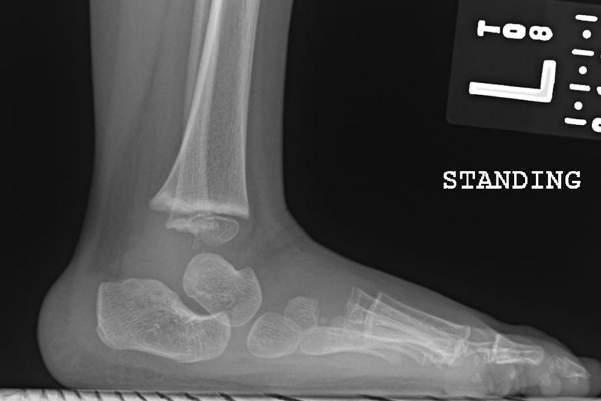
Finally, total correction of most talipes equinovarus may not be achievable. Some individuals will have some minimal residual heel varus and some adductus of the forefoot. However, the necessity for major reconstruction of joints has now been largely eliminated.
Dr. Harris is a Clinical Associate Professor in the Department of Orthopaedics and Rehabilitation at the Loyola Medical Center in Maywood, Ill. He is a Fellow of the American College of Foot and Ankle Surgeons.
References
- Mahan ST, Spencer SA, Kasser JR. Satisfactory patient-based outcomes after surgical treatment for idiopathic clubfoot: includes surgeon’s individualized technique. J Pediatr Orthop. 2014;34(6):631-8.
- Carroll NC. Clubfoot in the twentieth century: where we were and where we may be going in the twenty-first century. J Pediatr Orthop B. 2012;21(1):1-6.
- Van Gelder JH, van Ruiten AG, Visser JD, Maathuis PG. Long-term results of the posteromedial release in the treatment of idiopathic clubfoot. J Pediatr Orthop. 2010;30(7):700-4.
- Scher DM, Feldman DS, van Bosse HJ, Sala DA, Lehman WB. Predicting the need for tenotomy in the Ponseti method for correction of clubfeet. J Pediatr Orthop. 2004;24(4):349-52.
- MacNeille R, Hennrikus W, Stapinski B, Leonard G. A mini-open technique for Achilles tenotomy in infants with clubfoot. J Child Orthop. 2016;10(1):19-23.
- Pulak S, Swamy M. Treatment of idiopathic clubfoot by Ponseti technique of manipulation and serial plaster casting and its critical evaluation. Ethiop J Health Sci. 2012;22(2):77-84.
- Sharma S, Butt MF, Singh M, Sharma S. The posterior to anterior controlled technique of percutaneous Achilles tenotomy in the correction of idiopathic clubfoot: a technical report. J Pediatr Orthop B. 2013;22(3):249-51.
- Lebel E, Karasik M, Bernstein-Weyel M, et al. Achilles tenotomy as an office procedure: safety and efficacy as part of the Ponseti serial casting protocol for clubfoot. J Pediatr Orthop. 2012;32(4):412-5.
- Besselaar AT, Sakkers RJ, Schuppers HA, et al. Guideline on the diagnosis and treatment of primary idiopathic clubfoot. Acta Orthop. 2017:1-5.
- Jackson T, Jones A, Miller N, Georgopoulos G. Clubfoot and tethered cord syndrome: results of treatment with the Ponseti method. J Pediatr Orthop. 2017; epub Feb. 7.
- Lebel E, Weinberg E, Berenstein-Weyel TM, Bromiker R. Early application of the Ponseti casting technique for clubfoot correction in sick infants at the neonatal intensive care unit. J Pediatr Orthop B. 2017;26(2):108-111.
- Radler C, Mindler GT. [Pediatric clubfoot : Treatment of recurrence]. Orthopade. 2016;45(10):909-24.
- Parsa A, Moghadam MH, Jamshidi MH. Relapsing and residual clubfoot deformities after the application of the Ponseti method: a contemporary review. Arch Bone Jt Surg. 2014;2(1):7-10.
- Saetersdal C, Fevang JM, Bjorlykke JA, Engesaeter LB. Ponseti method compared to previous treatment of clubfoot in Norway. A multicenter study of 205 children followed for 8-11 years. J Child Orthop. 2016;10(5):445-52.
- Thompson GH, Simons GW. Congenital talipes equinovarus (clubfeet) and metatarsus adductus. In: Drennan JC (ed.). The Child’s Foot and Ankle. Raven Press, New York, 1992, pp. 97-133.
- Matar HE, Beirne P, Bruce CE, Garg NK. Treatment of complex idiopathic clubfoot using the modified Ponseti method: up to 11 years follow-up. J Pediatr Orthop B. 2017;26(2):137-142.
- Gurnett CA, Boehm S, Connolly A, Reimschisel T, Dobbs MB. Impact of congenital talipes equinovarus etiology on treatment outcomes. Dev Med Child Neurol. 2008;50(7):498-502.
- Hosseinzadeh P, Steiner RB, Hayes CB, et al. Initial correction predicts the need for secondary Achilles tendon procedures in patients with idiopathic clubfoot treated with Ponseti casting. J Pediatr Orthop. 2016;36(1):80-3.
- Alvarado DM, Buchan JG, Frick SL, et al. Copy number analysis of 413 isolated talipes equinovarus patients suggests role for transcriptional regulators of early limb development. Eur J Hum Genet. 2013;21(4):373-80.
- Alvarado DM, Aferol H, McCall K, et al. Familial isolated clubfoot is associated with recurrent chromosome 17q23.1q23.2 microduplications containing TBX4. Am J Hum Genet. 2010;87(1):154-60.
- Dobbs MB, Gurnett CA. Genetics of clubfoot. J Pediatr Orthop B. 2012;21(1):7-9.
- Shyy W, Dietz F, Dobbs MB, Sheffield VC, Morcuende JA. Evaluation of CAND2 and WNT7a as candidate genes for congenital idiopathic clubfoot. Clin Orthop Relat Res. 2009;467(5):1201-5.
- Cao D, Jin C, Ren M, Lin C, Zhang X, Zhao N. The expression of Gli3, regulated by HOXD13, may play a role in idiopathic congenital talipes equinovarus. BMC Musculoskelet Disord. 2009;10:142.
- Matar HE, Beirne P, Garg NK. Effectiveness of the Ponseti method for treating clubfoot associated with myelomeningocele: 3-9 years follow-up. J Pediatr Orthop B. 2017;26(2):133-136.
- Matar HE, Beirne P, Garg N. The effectiveness of the Ponseti method for treating clubfoot associated with arthrogryposis: up to 8 years follow-up. J Child Orthop. 2017;10(1):15-8.
- Kowalczyk B, Felus J. Ponseti casting and Achilles release versus classic casting and soft tissue releases for the initial treatment of arthrogrypotic clubfeet. Foot Ankle Int. 2015;36(9):1072-7.
- Morcuende JA, Dobbs MB, Frick SL. Results of the Ponseti method in patients with clubfoot associated with arthrogryposis. Iowa Orthop J. 2008;28:22-6.
- Garg S, Dobbs MB. Use of the Ponseti method for recurrent clubfoot following posteromedial release. Indian J Orthop. 2008;42(1):68-72.
- Radler C, Suda R, Manner HM, Grill F. [Early results of the Ponseti method for the treatment of idiopathic clubfoot]. Z Orthop Ihre Grenzgeb. 2006;144(1):80-6.
- Miller NH, Carry PM, Mark BJ, Engelman GH, Georgopoulos G, Graham S, et al. Does strict adherence to the Ponseti method improve isolated clubfoot treatment outcomes? A two-institution review. Clin Orthop Relat Res. 2016;474(1):237-43.
- Hayes CB, Murr KA, Muchow RD, Iwinski HJ, Talwalkar VR, Walker JL, et al. Pain and overcorrection in clubfeet treated by Ponseti method. J Pediatr Orthop B. 2017; epub Feb. 24.
- Hosseinzadeh P, Kelly DM, Zionts LE. Management of the relapsed clubfoot following treatment using the Ponseti method. J Am Acad Orthop Surg. 2017;25(3):195-203.
- Hosseinzadeh P, Kiebzak GM, Dolan L, Zionts LE, Morcuende J. Management of clubfoot relapses with the Ponseti method: results of a survey of the POSNA Members. J Pediatr Orthop. 2017; epub Feb. 7.
- Maripuri SN, Gallacher PD, Bridgens J, Kuiper JH, Kiely NT. Ponseti casting for club foot - above- or below-knee?: A prospective randomised clinical trial. Bone Joint J. 2013;95-B(11):1570-4.
- Karami M, Dehghan P, Moshiri F, Shamami MS. Effect of unintentional partial Achilles tenotomy on Ponseti clubfoot management outcomes. J Pediatr Orthop B. 2015;24(1):1-5.
- Agarwal A, Kumar A, Shaharyar A, Mishra M. The problems encountered in a CTEV clinic: can better casting and bracing be accomplished? Foot Ankle Spec. 2016; epub Sept 7.
- Seegmiller L, Burmeister R, Paulsen-Miller M, Morcuende J. Bracing in Ponseti clubfoot treatment: improving parental adherence through an innovative health education intervention. Orthop Nurs. 2016;35(2):92-7; quiz 98-9.
- Aydin BK, Sofu H, Senaran H, et al. Treatment of clubfoot with Ponseti method using semirigid synthetic softcast. Medicine (Baltimore.) 2015;94(47):e2072.
- Liu Y, Zhao D, Zhao L, Li H, Yang X. Congenital Clubfoot: Early Recognition and Conservative Management for Preventing Late Disabilities. Indian J Pediatr. 2016;83(11):1266-1274.
- Zhao D, Liu J, Zhao L, Wu Z. Relapse of clubfoot after treatment with the Ponseti method and the function of the foot abduction orthosis. Clin Orthop Surg. 2014;6(3):245-52.
- Morin ML, Hoopes DM, Szalay EA. Positive communication paradigm decreases early recurrence in clubfoot treatment. J Pediatr Orthop. 2014;34(2):219-22.
- Radler C. The Ponseti method for the treatment of congenital club foot: review of the current literature and treatment recommendations. Int Orthop. 2013;37(9):1747-53.
- Hemo Y, Segev E, Yavor A, et al. The influence of brace type on the success rate of the Ponseti treatment protocol for idiopathic clubfoot. J Child Orthop. 2011;5(2):115-9.
- Zionts LE, Dietz FR. Bracing following correction of idiopathic clubfoot using the Ponseti method. J Am Acad Orthop Surg. 2010;18(8):486-93.
- Goldstein RY, Seehausen DA, Chu A, Sala DA, Lehman WB. Predicting the need for surgical intervention in patients with idiopathic clubfoot. J Pediatr Orthop. 2015;35(4):395-402.
- Dobbs MB, Rudzki JR, Purcell DB, et al. Factors predictive of outcome after use of the Ponseti method for the treatment of idiopathic clubfeet. J Bone Joint Surg Am. 2004;86-A(1):22-7.
- Goksan SB, Bilgili F, Eren I, Bursali A, Koc E. Factors affecting adherence with foot abduction orthosis following Ponseti method. Acta Orthop Traumatol Turc. 2015;49(6):620-6.
- Memon I, Bhatti A, Ali P, Mahmood K. Difficulties in maintenance of clubfoot abduction brace and solutions - maintenance of clubfoot abduction brace, locks and keys. J Pak Med Assoc. 2014;64(12 Suppl 2):S70-5.
- Bhaskar A, Patni P. Classification of relapse pattern in clubfoot treated with Ponseti technique. Indian J Orthop. 2013;47(4):370-6.
- Lovell ME, Morcuende JA. Neuromuscular disease as the cause of late clubfoot relapses: report of 4 cases. Iowa Orthop J. 2007;27:82-4.
- Zhao D, Li H, Zhao L, Liu J, Wu Z, Jin F. Results of clubfoot management using the Ponseti method: do the details matter? A systematic review. Clin Orthop Relat Res. 2014;472(4):1329-36.
- Sugandhavesa N, Cheewawattanachai C, Luevitoonvechkij S, Khunsree S. Results of shortened program of Ponseti technique for congenital clubfoot. J Med Assoc Thai. 2015;98(1):88-92.
- Xu RJ. A modified Ponseti method for the treatment of idiopathic clubfoot: a preliminary report. J Pediatr Orthop. 2011;31(3):317-9.
- Harnett P, Freeman R, Harrison WJ, Brown LC, Beckles V. An accelerated Ponseti versus the standard Ponseti method: a prospective randomised controlled trial. J Bone Joint Surg Br. 2011;93(3):404-8.
- Hui C, Joughin E, Nettel-Aguirre A, Goldstein S, Harder J, Kiefer G, et al. Comparison of cast materials for the treatment of congenital idiopathic clubfoot using the Ponseti method: a prospective randomized controlled trial. Can J Surg. 2014;57(4):247-53.
- Gintautiene J, Cekanauskas E, Barauskas V, Zalinkevicius R. Comparison of the Ponseti method versus early tibialis anterior tendon transfer for idiopathic clubfoot: A prospective randomized study. Medicina (Kaunas). 2016;52(3):163-70.
- Grigoriou E, Abol Oyoun N, Kushare I, et al. Comparative results of percutaneous Achilles tenotomy to combined open Achilles tenotomy with posterior capsulotomy in the correction of equinus deformity in congenital talipes equinovarus. Int Orthop. 2015;39(4):721-5.
- Kumar MN, Gopalakrishna C. Modified Ponseti method of management of neonatal club feet. Acta Orthop Belg. 2012;78(2):210-5.
- Iltar S, Uysal M, Alemdaroglu KB, et al. Treatment of clubfoot with the Ponseti method: should we begin casting in the newborn period or later? J Foot Ankle Surg. 2010;49(5):426-31.
- Bashi RH, Baghdadi T, Shirazi MR, Abdi R, Aslani H. Modified Ponseti method of treatment for correction of neglected clubfoot in older children and adolescents--a preliminary report. J Pediatr Orthop B. 2016;25(2):99-103.
- Faizan M, Jilani LZ, Abbas M, Zahid M, Asif N. Management of Idiopathic clubfoot by ponseti technique in children presenting after one year of age. J Foot Ankle Surg. 2015;54(5):967-72.
- Yagmurlu MF, Ermis MN, Akdeniz HE, Kesin E, Karakas ES. Ponseti management of clubfoot after walking age. Pediatr Int. 2011;53(1):85-9.
- Sinha A, Mehtani A, Sud A, et al. Evaluation of Ponseti method in neglected clubfoot. Indian J Orthop. 2016;50(5):529-535.
- Banskota B, Banskota AK, Regmi R, et al. The Ponseti method in the treatment of children with idiopathic clubfoot presenting between five and ten years of age. Bone Joint J. 2013;95-B(12):1721-5.


