

ADVERTISEMENT
How To Address Subtalar Joint Instability
Emphasizing careful assessment of the etiology and underlying contributing factor to subtalar joint instability, this author reviews pertinent conservative and surgical options for treating this conditin in children and adults. There are two forms of subtalar joint instability. It has been recognized as a cause of ankle symptoms secondary to ligamentous injury or laxity of the subtalar joint. This contributes to instability of the ankle as well. Studies show that sectioning of the calcaneal fibular ligament and cervical ligament of the subtalar joint can lead to as much as a 7 mm increase in talar tilt. Surgical repairs to correct lateral ankle instability should include repair or substitution of the calcaneofibular ligament if subtalar instability is a consideration. The other form of subtalar instability is associated with an excessive range of motion with or without pathology. It can be a process of increased motion that leads to minor pathology and muscular fatigue. Alternatively, it may cause a more complex situation of excessive pronatory motion, leading to collapse of the midtarsal joint and global mechanical problems for the foot and lower extremity. For the purposes of this article, I will consider subtalar joint instability as excessive subtalar joint motion or a position that leads to pain and subjective or objective complaints for a patient.
Treating Excessive Subtalar Joint Motion In Children
One may appreciate excessive subtalar joint motion early in a child’s life. Often young children will come in with their parents and present with markedly flatfeet and the majority of the collapse originates from the subtalar joint. More often than not, these patients do not have pain but there is a fear of the future ramifications of this position. A full evaluation of the possible pathological etiologies of this condition is necessary. The majority of the children will present these findings as their normal musculoskeletal position. It is debatable whether one should treat a painless/ unstable foot in this situation. Certainly, there are situations in which there is accompanying pain or when findings are so clear that one must institute treatment. However, in the majority of cases involving excessive subtalar joint motion in children, it has been our experience that it is usually best to leave these patients untreated if they are asymptomatic. Over the years of treating professional athletes, it has been our observation that many of the fastest runners and highest jumpers have significant flatfeet that were likely present since childhood. It is hard for us to justify treatment in a child without pain considering the level of achievement of unstable subtalar joints in the professional athletic population. In children who have pain associated with instability of the subtalar joint, utilizing a functional orthotic device or UCBL device can have profound effects on the foot. Children who suffer from pain associated with subtalar joint instability will frequently complain of vague foot/ankle pain, leg pain, “growing pain” and fatiguing quickly when walking or running. By stabilizing the subtalar joint with an external supportive device, clinicians can facilitate more efficient function of the lower extremity and allow for a return to normal activities with reduced pain. It has been our experience that even with the most unstable subtalar joints in children, one can facilitate control with orthotic devices and that nearly all of these patients should receive this treatment alternative initially. However, in some situations, the subtalar joint is too unstable to be controlled by an external orthotic device and one must consider more aggressive measures. When orthotics fail and pain and problems persist, subtalar joint arthroereisis becomes a viable alternative. Over the years, podiatric physicians have frequently utilized arthroereisis in these situations. Few strong, scientific, published reports support the benefits of arthroereisis but those who have the greatest experience with the procedure laud its utility. We have found that absorbable materials may lead to equal success in controlling an unstable subtalar joint without the fear of long-term problems of a metallic implant causing subtalar joint irritation and erosion. Based on the anecdotal experience of those who have had to remove painful metallic arthroereisis implants and reported maintenance of correction, we started using absorbable materials for implantation and have had consistent success. Giannini has reported similar findings on a large series of absorbable implant cases in children.1
When Adults Present With Subtalar Joint Instability
It is more common to see adults present with pain and problems associated with subtalar joint instability. In clinicial scenarios that involve minor instability, patients will present with an array of foot and ankle related problems. However, subtalar joint instability can also lead to orthopedic problems affecting the knees, hips and lower back. When the subtalar joint is unstable, the biomechanical effects can be tremendous. The midtarsal joint becomes more unstable and forefoot and rearfoot problems become more pronounced. Common foot and ankle presentations of subtalar instability are posterior tibial tendonitis, anterior tibial tendonitis, plantar fasciitis and forefoot pain commonly associated with lesser metatarsal head overload. Initial treatment for these entities includes improved shoe gear, supportive devices and taping and bracing techniques. Often, the problems of these patients require more permanent support and control, and custom orthotics become necessary. There is a wide array of materials one can utilize for the fabrication of orthotics but it has been our experience that softer orthotics with deep heel seats and medial and lateral flanges are most effective. Not only do these orthotics facilitate comfortable control of these entities, they make the use of forefoot accommodations more tolerable. Additionally, we have found that computer scanning of the foot provides a consistent impression of the foot equal to that of more traditional impression techniques. The computer scanning allows for streamlining of our custom orthotic fabricating process and has tremendously reduced office overhead and costs. When rearfoot and ankle tendon pathologies are present, bracing techniques can have benefits. In the case of early stage tendinitis, there is a wide variety of braces available to control subtalar joint motion in order to reduce the stress on the tendons that are functionally trying to control subtalar joint motion. We prefer to utilize braces that control the subtalar joints while still allowing for somewhat normal shoe wear. The use of a foot/ankle brace with straps is a very effective tool to control these problems and still permit normal shoe wear. Patients can easily utilize these braces in sports activities. Studies have shown an equal benefit of braces to sports taping techniques with better cost effectiveness because of the ability to reuse the brace.2
Pertinent Pearls On Managing Severe Or Chronic Instability
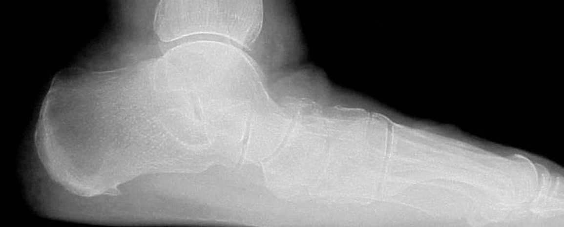
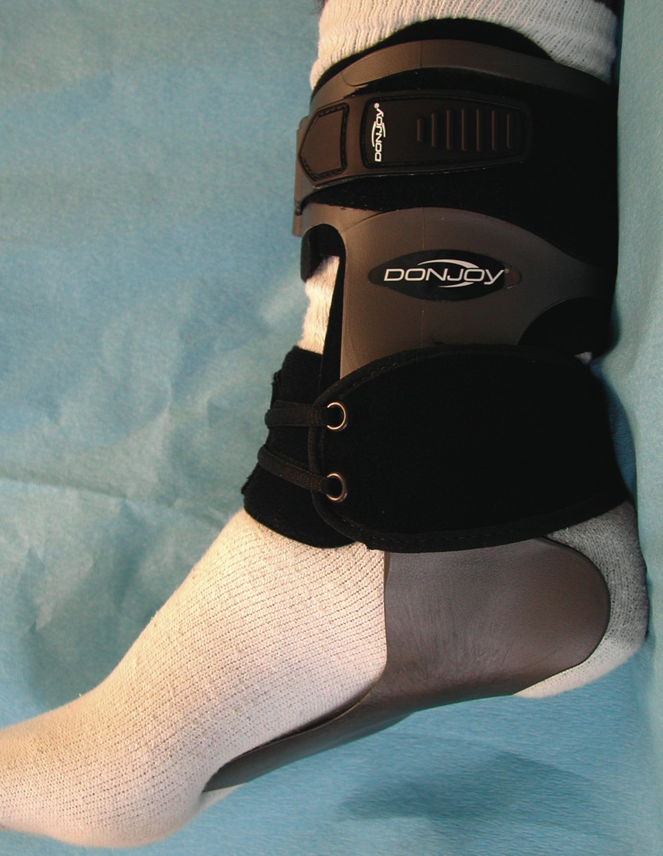
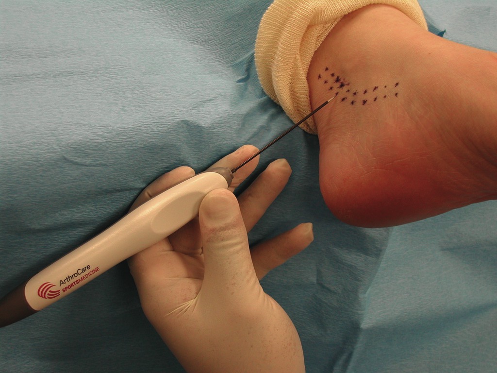
Unfortunately, in many situations, pathologies are too severe or too chronic to be controlled with orthotics and/or simple bracing techniques. In those situations, more aggressive bracing techniques can prove valuable. For patients with uncontrolled subtalar instability, the Velocity Brace (DJO) has become a mainstay in our practice. The Velocity Brace is a very supportive foot/ankle brace with a low profile. There is a plastic foot plate that one can heat mold to a patient’s foot to offer customizable comfort and control. The brace also offers the benefit of crossing the subtalar and ankle joint to control triplane instability. The brace’s comfort and low profile allow for high patient compliance with this device. & In the situation that off-the-shelf braces do not provide enough support for severely unstable subtalar joints, full custom braces can be the answer. A modified ankle foot orthosis (AFO) has become the standard for these situations. At our in-house brace laboratory, The Footcare Depot, we offer a custom-made, leather gauntlet modified AFO for the most severe problems. This is a useful device for severe deformity or patients who are not appropriate surgical candidates. & Sometimes orthotic and bracing techniques are not completely effective in relieving pain or not acceptable to patient needs. When treating these patients, one will often note MRI changes, which frequently occur in the tibialis posterior tendon with longitudinal tears or thickening of the tendon. We have been successful in utilizing minimally invasive techniques to surgically address these conditions. When the patient does not have severe pathology to the posterior tibial tendon, we will often perform a percutaneous microtenotomy with coblation in combination with an absorbable subtalar arthroereisis. This minimally invasive combination allows for reduced recovery time with a supportive walking boot for two to three weeks followed by a return to normal shoe gear. After undergoing this combination of surgical procedures, most patients are able to return to full activities eight to 10 weeks postoperatively. & When it comes to the most severe and chronic subtalar joint instability, it is common to find patients with a complete loss of function of the posterior tibial tendon. Subtalar joint instability can cause such stress on the posterior tibial tendon that attenuation and/or rupture of the tendon can occur, leading to greater instability of the subtalar joint. When this happens, patients will complain of a progressive collapse of the arch, foot or ankle, which frequently occurs over a period of six months. Pain may or may not be present at the medial foot and ankle. While pain is usually present over the course of the tibialis posterior tendon, it can commonly be present at the lateral subtalar joint due to excessive collapse of the subtalar joint causing lateral impingement. The physical examination will reveal severe collapse of one foot with an inability to rise up on the toes of the problematic foot. Employing diagnostic ultrasound or MRI can help confirm that diagnosis. These patients usually need aggressive care with either a leather gauntlet modified AFO or corrective foot surgery.
Exploring Surgical Options For Tibialis Posterior Dysfunction Due To STJ Instability
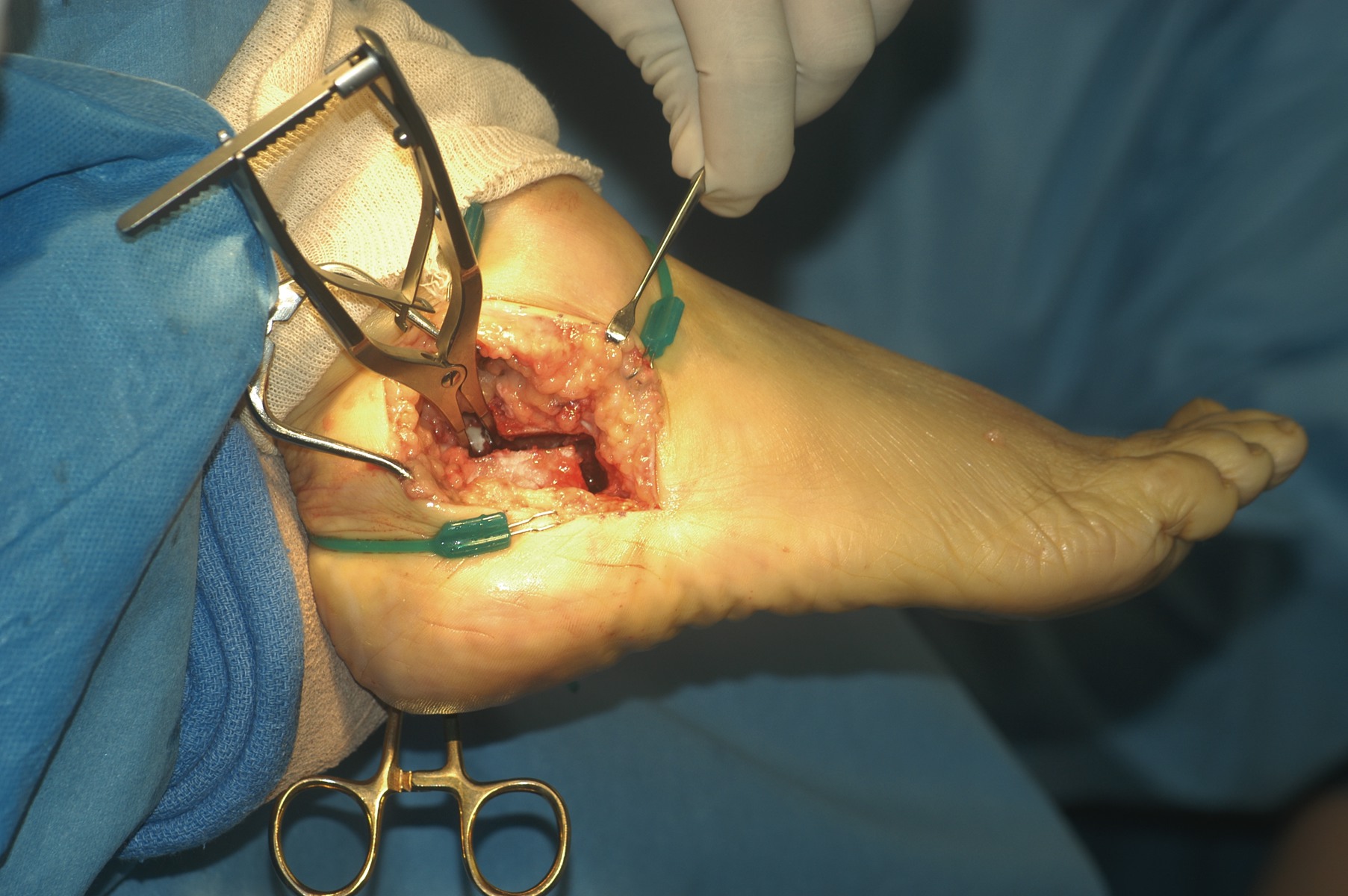
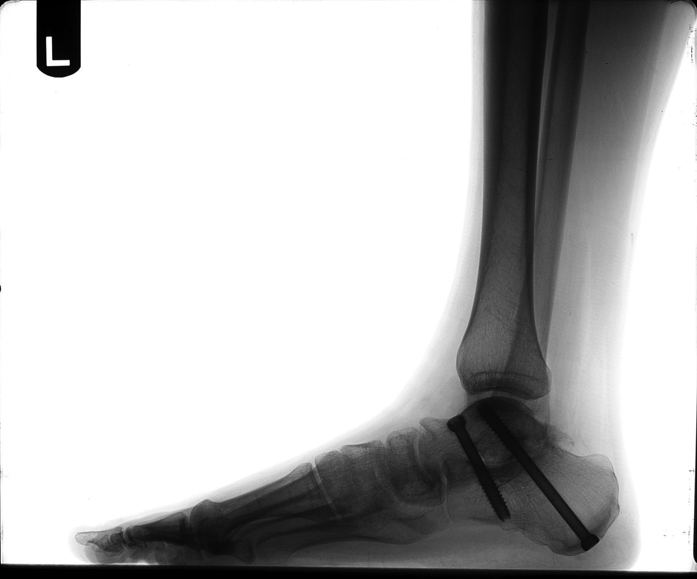
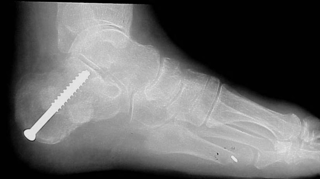
There are many surgical techniques for the treatment of tibialis posterior dysfunction due to an unstable subtalar joint. We prefer combining osseous and soft tissue procedures to correct the problem. Over the last six years, we have utilized the calcaneal Scarf to correct the rearfoot/subtalar position. The calcaneal Scarf allows accurate medial translation of the calcaneus while lengthening it in one osteotomy. The orientation of the osteotomy has increased stability over other calcaneal osteotomy options, allowing for ease of fixation and an earlier return to weightbearing. One benefit of this procedure is that it positions the subtalar joint from a valgus position into a more rectus alignment, thus reducing instability of the subtalar joint. This procedure also facilitates correction of transverse plane deformities. In the instance of severe instability that is not completely reduced by the calcaneal Scarf, we will augment our correction with an absorbable arthroereisis to further stabilize subtalar joint motion. & Correcting the weakened or absent tibialis posterior tendon is another crucial element to successful surgical outcomes. Podiatric surgeons commonly utilize a tendon transfer (flexor digitorum longus) or a tenodesis with the Cobb procedure (tibialis anterior). We prefer the Cobb procedure. However, in scenarios when one can avoid performing a tendon transfer, it is our preference to utilize the OrthoAdapt collagen scaffold (Pegasus Biologics). This material allows for reconstruction of the tibialis posterior tendon without sacrifice or insult to another tendon. With its cross-linked nature, the Orthoadapt is strong enough to maintain the correction. Its inherent collagen matrix also allows tissue in-growth, which ultimately will transition into normal host tendon tissue. In the most severe and longstanding cases of subtalar joint instability, arthritis of the subtalar joint may result. When bracing techniques fail in these situations, joint destructive procedures become an alternative. Studies have shown that surgeons can reduce 70 percent of subtalar joint motion by fusing the talonavicular joint. However, we prefer directly addressing the subtalar joint via an arthrodesis. & Arthrodesis of the subtalar joint will allow complete stability of the subtalar component of the rearfoot complex and positioning of the subtalar joint in optimal alignment. While fusion is never the preferred approach to any joint, it becomes necessary in many cases and can often lead to superior outcomes.
In Conclusion
We deal with subtalar joint instability every day in our podiatric practices. Careful assessment of the etiology and underlying contributions is paramount to successful treatment. Appropriate uses of the tools available to us can produce positive results for the patients, their families and the physician. Proper selection of techniques and intervention are crucial to optimal outcomes. Dr. Weil is the Fellowship Director of the Weil Foot and Ankle Institute in Des Plaines, Ill. He is a Fellow of the American College of Foot and Ankle Surgeons.
References:
References 1. Giannini S, et al. Operative treatment of flatfoot with talocalcaneal coalition. Clin Orthop Rel Res. 411:178-87, June 2003. 2. Mickel TJ, Bottoni CR, Tsuji G, et. al. Prophylactic bracing versus taping for the prevention of ankle sprains in high school athletes: a prospective, randomized trial. J Foot Ankle Surg 45(6):360-365.


