

ADVERTISEMENT
How To Detect Pediatric Osteomyelitis
Although both acute hematogenous osteomyelitis (AHO) and chronic recurrent multifocal osteomyelitis (CRMO) are somewhat uncommon, differentiating between the two can be tricky. Having a strong grasp of the etiology and presentation of these conditions can go a long way toward preventing a delayed or inappropriate diagnosis, which could be harmful for pediatric patients. Pediatric hematogenous osteomyelitis may occur secondary to a traumatic injury and/or an acquired illness or other immunosuppressive condition. Acute hematogenous osteomyelitis reportedly affects one in 5,000 children in the United States under the age of 13 with half of all cases occurring in children under 5.1-3 The condition can be quite dangerous in prognosis if the diagnosis is delayed. Therefore, it is essential to be able to differentiate between AHO and other conditions. Chronic recurrent multifocal osteomyelitis is a separate and rare disorder that affects children and teenagers. The condition is an inflammatory disorder of unknown etiology that involves different osseous sites and has some association with psoriasis and palmoplantar pustulosis. While antibiotic therapy usually works for AHO, one would primarily treat CRMO with antiinflammatories. That said, let us take a closer look at each condition.
Understanding The Presentation And Pathophysiology Of AHO
Acute hematogenous osteomyelitis in children is an uncommon but not rare systemic disease that occurs primarily in the metaphysis of long bones.1 Morbidity mostly occurs via delays in diagnosis that may be due to negative culture results and/or a poor understanding of the pathophysiology of bone infections that can lead to mismanagement of the patient.1 The earliest presentation has no findings other than local tenderness and an elevated erythrocyte sedimentation rate (ESR).4 Children may acquire osteomyelitis via three sources. The most common sources include contiguous or soft tissue sources (i.e., excoriations, abrasions, insect or animal bites), puncture wounds and hematogenous spread. A history of trauma or other insinuating event to the body can potentiate the infestation of bacteria into the bloodstream. Guler, et. al., sought to establish the role of blunt trauma without fracture or obvious wound as a risk factor in children admitted for community-acquired Staph septicemia.5 They showed that skeletal involvement was multifocal and more frequent in traumatized, older patients.5 The period between the symptoms of infection and diagnosis was also shorter in traumatized patients and included 12 (46 percent) of 26 septicemic patients.5 Viruses or bacterial infections, such as an upper respiratory infection, pharyngitis and otitis media, can also lead to an infection in the bloodstream, and potentially affect the bone and joints. The patient history and physical exam findings may be sensitive but they are rarely specific. Typically, fever, pain and limited use of an affected extremity are the most common symptoms whereas constitutional symptoms are less common. Older children will have even more subtle presentations of symptoms. Arthritic conditions such as juvenile rheumatoid arthritis can make joints more susceptible to invasion by organisms. Accordingly, one should rule out such conditions during a routine screening if a child presents with any joint or limb pain. The pathophysiology of AHO begins with an infection entering the bone into the metaphysis through the haversian system. This can lead to abscess formation as the periosteum becomes elevated and the infection invades the subperiosteal space. With the exception of neonates and infants, involvement of two or more sites is uncommon as AHO is usually a unilateral condition.1 Neonates and infants have a more vascular nature of the physis with the physis becoming more avascular by 15 to 18 months of age, preventing further spread and localizing infections to the metaphysis.1 Since blood vessels cross the physis until infants are 15 to 18 months of age, the potential for developing epiphyseal osteomyelitis as well as septic arthritis increases during this time period. The estimated number of neonatal osteomyelitis cases is one to three infants per 1,000 intensive care/nursery admissions.3 Although AHO is more common in the metaphysis, Kramer reported a case of an 11-year-old child with epiphyseal involvement.6 At that time, there were no other reported findings showing epiphyseal involvement.
Pertinent Diagnostic Pearls
While plain radiographs may show joint effusions and a loss of soft tissue planes, there may not be an immediate indication of bone infection.7 It may take 10 to 14 days to show 30 to 50 percent of bone mineral loss before osteomyelitic changes are evident.1 A differential diagnosis for radiographic bony lesions should include osteomyelitis, leukemia, round cell carcinomas, Ewing’s sarcoma, metastatic neuroblastomas, eosinophilic granulomas, histiocytosis X and tuberculosis. If radiographs are normal and one suspects osteomyelitis, proceed to obtain technetium-99m bone scans. However, keep in mind that while this imaging is sensitive for certain bony conditions, it is not specific. Potential pediatric conditions that could show an increase in tracer uptake include osteomyelitis, reflex sympathetic dystrophy, arthritidies, fractures, tumors and active growth plates. Sensitivity can range from 84 to 100 percent for detection of osteomyelitis and 70 to 96 percent for specificity.1,8 If Tc-99m appears normal and one still suspects osteomyelitis, indium 111-tagged white blood cell (WBC) scans can be helpful. Ultrasound is great in detecting intraarticular, soft tissue and subperiosteal fluid collections. Benefits of using this modality include a lower cost and ready availability. The non-invasive nature of ultrasound and the lack of a need for sedation make the diagnostic imaging a less traumatic experience for the child.1 When it comes to a child with possible AHO or suspected septic arthritis, one should order the following serologic studies: complete blood count (CBC) with differential, arthritic panel, peripheral smear, ESR, C-reactive protein (CRP) and blood cultures. However, be aware that blood work for AHO does not always indicate an infection either. One will see an elevated WBC in approximately 31 to 40 percent of the pediatric population and an elevated ESR in 91 percent of the patients.1,9-11 Blood cultures are positive in approximately 30 to 60 percent of cases of AHO in children.2 Clinicians should evaluate a peripheral smear to rule out any underlying malignant disease such as leukemia.1
What Treatments Are Effective for AHO?
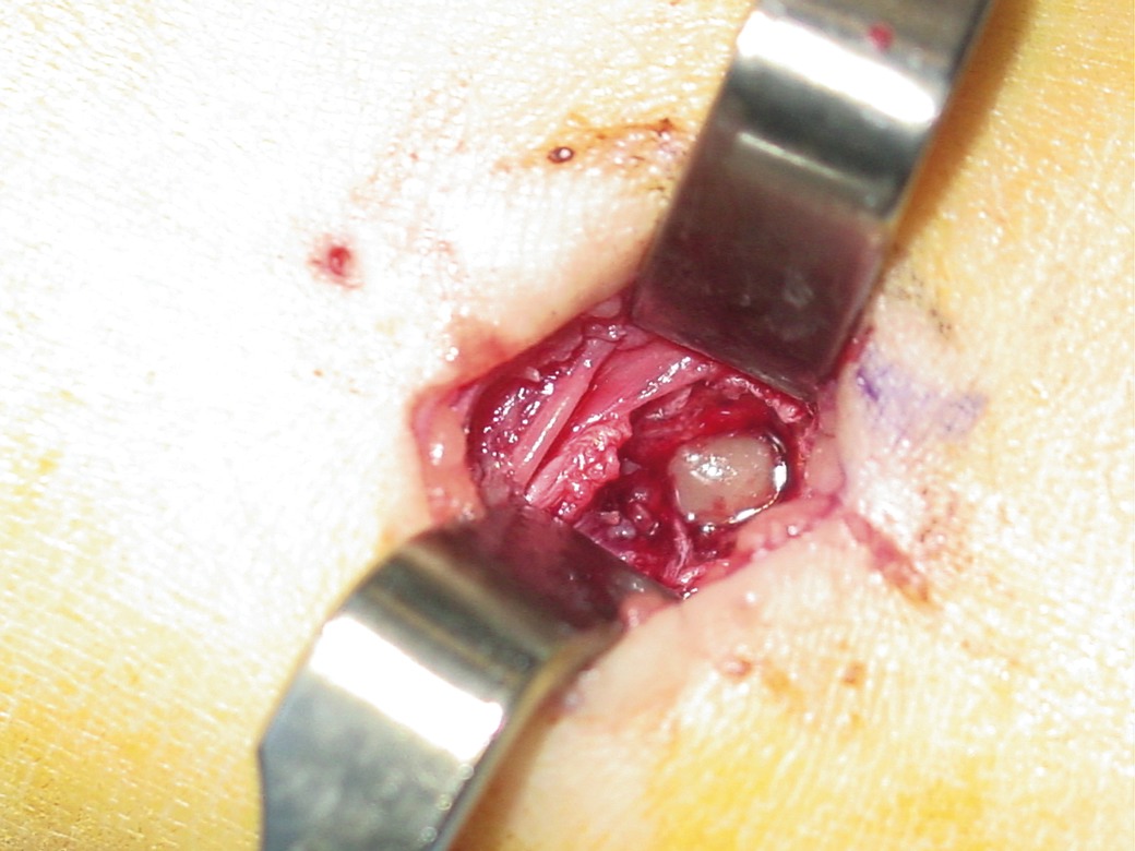
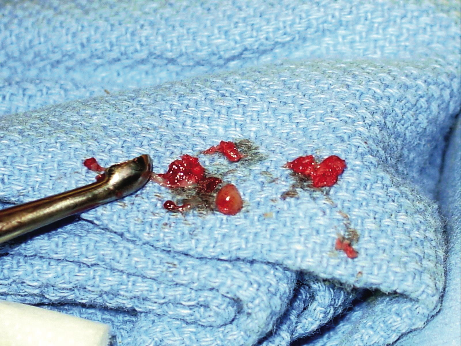
The treatment for AHO is usually antibiotic therapy and/or surgical intervention, which includes incision and drainage of abscesses, as well as debridement of infected, devitalized tissue and bone. One should begin empiric treatment by targeting gram-positive organisms. Researchers found that Staphylococcus aureus is the most common organism in 89 percent of these infections.1 Streptococcus pneumoniae, Group A Streptococcus and coagulase negative Staphylococcus are more age- and disease-specific.1 Researchers have found group B streptococci in greater frequency in neonates.1 One should ultimately tailor antibiotic therapy to target the culture results of soft tissue and bone specimens. Recent studies suggest using a sequential intravenous-oral antibiotic regimen.3 Again, it is important to differentiate between AHO and CRMO as CRMO does not respond to antibiotics but usually does respond to antiinflammatory medication.12
A Guide To The Etiology And Presentation Of CRMO
Chronic recurrent multifocal osteomyelitis is a systemic, non-purulent disorder that is characterized by having an insidious onset with local swelling and pain.13-18 The disorder has also been known as chronic multifocal symmetric osteomyelitis, chronic symmetric osteomyelitis, chronic multifocal cleido-metaphyseal osteomyelitis and subacute symmetric osteomyelitis.19,20 Chronic recurrent multifocal osteomyelitis, which clinicians usually diagnose in patients around the age of 2, is benign and self-limiting. Researchers have noted that CRMO has a long chronic course with recurrences and remissions that can last up to six to 10 years.17-19 There has been no racial predisposition and females are affected twice as often as males with half the cases involving children 10 years and younger.21 It is a distinct clinical entity and differentiating it from subacute bacterial osteomyelitis and AHO is difficult.20 Giedion, et. al., first described the condition in 1972 and Girschicke, et. al., called CRMO an inflammatory disorder of unknown etiology that involves different osseous sites, primarily the metaphysis of long bones and pelvic bones with a widely reported incidence involving the knees and ankles.12,16,22-25 Of the 50 cases Van Howe reported, the tibia was the most common location with 37 lesions and there were 16 lesions in the foot.19 A symmetrical, recurrent and multifocal pattern is the usual presentation of this condition. However, although initial reports showed symmetry, this has not been a consistent finding in subsequent reports.19 The child may present with pain, stiffness and fever. He or she may be afebrile, limping, unable to bear weight on affected limbs and one may notice swelling of bones and joints that lasts a few months in duration. The diagnosis of CRMO becomes one of exclusion due to the lack of pathognomonic findings.23,24 With this condition, there are no consistent laboratory findings with the exception of an elevated erythrocyte sedimentation rate (ESR).24 Indeed, this diagnosis can be very challenging. After documenting the initial presentation and work-up, the clinician may commonly misdiagnose the problem as AHO, rheumatic fever, juvenile rheumatoid arthritis, subacute bacterial osteomyelitis, viral polyarthritis, osteochondrosis juvenilis and enthesiopathy. The pathogenesis is unknown but it has been associated with SAPHO (synovitis, acne, pustulosis, hyperostosis, osteitis) syndrome.26,27 SAPHO and CRMO have similar features and have a generally accepted association.27 One may first recognize the pathogenesis as an enthesiopathy that progresses to the osseous portion of the tendon and produces a non-bacterial chronic osteomyelitis.18 There has also been an inconsistent association between CRMO and cutaneous disorders, including acne fulminans, psoriasis and palmoplantar pustulosis.13,14,18,21 Van Howe found a 20 percent incidence of CRMO involving patients who developed a pustular rash on their palms and soles.19 Anhurst found involvement of the heel and in-step of the feet.28 Schilling found a 25 percent association of children and 50 percent of adults having a relationship of CRMO and pustulous dermatosis.16 Majeed, et. al., found an association between chronic dyserythropoietic anemia and CRMO.29 They described two patients afflicted with the autosomal recessive syndrome. Coinde, et. al., refer to CRMO as an osteitis of unknown etiology that is mistaken as osteomyelitis.13 Giedion speculated about an autoimmune etiology but Bjorksten and Boquist thought this was unlikely.22,25 They found an accumulation of PMNs, lymphocytes, plasma cells, fibrosis and osteosclerosis around inflammatory infiltrates, representing a clinical entity of unknown etiology.25 Speer came up with several possible etiologies for CRMO. These include a chronic or “burned-out” osteomyelitis; a non-infectious process with regional metaphyseal influences; a viral infection of bone; or an organism with fastidious growth requirements not met in standard culture techniques.30
What Does The Literature Reveal About Diagnosing And Treating CRMO?
When it comes to CRMO, radiographic findings include osteolysis, expansion or periosteal reaction, sclerosis and onion skinning.19,31 Bone scans are ideal for identifying the multifocality of CRMO and one may subsequently use radiographs and CT scans to evaluate these focal areas.19 Clinical and radiologic differential diagnosis may include Ewing’s sarcoma, Langerhans cell histiocytosis (mainly tuberculosis), hematolymphoid malignancy and metastatic neuroblastoma.32 Along with the presenting factors and the presence of osteolytic lesions on X-rays, the clinician should consider the possibility of multiple inflammatory and neoplastic processes including hematogenous osteomyelitis, Brodie’s abscess, subacute osteomyelitis histiocytosis X, acute types of leukemia, rhabdomyosarcoma and metastatic neuroblastoma.19,33 Overall, CRMO is a great clinical and radiological mimic that merits the physician’s recognition.32 Biopsies are required to rule out an infectious bacterial osteomyelitis or tumoral process yet the histiopathic examinations in CRMO may not always show bacterial infestation.27 Researchers recommend open biopsies over percutaneous biopsies due to the high potential of contaminants.19 In a study of 17 cases of pediatric osteomyelitis by Coinde, et. al., histopathological exams showed only mild inflammatory, non-specific changes and cultures were negative.13 An article by Chow, et. al., also demonstrated poor correlations between histologic and clinical features.32 Van Howe reported a case of an 8-year-old girl who had toe pain for several weeks. Employing bone scan, he discovered multiple areas of other involved asymptomatic sites.19 The biopsy was negative and although the patient took no antibiotics or antiinflammatories, she recovered uneventfully. Since CRMO is essentially self-limiting, one would usually emphasize supportive treatment with antiinflammatory therapy and active rehabilitation after a period of prolonged rest.19 Studies have shown that children taking antibiotics to treat this condition did as well as those who did not. Duffy, et. al., showed some symptomatic benefit with intravenous bisphosphonates.31
In Conclusion
Differentiating between AHO and CRMO may be difficult. Both CRMO and AHO can present with negative culture results, normal blood work and an increase in the ESR. Histopathologic features alone may not provide conclusive evidence. In order to make a definitive diagnosis, one should examine the entire clinical picture, taking X-rays, bone scans and MRI, bacterial cultures and histopathologic analysis into account as well as including a multispecialty team approach. Ultrasound is also a quick, inexpensive diagnostic tool for localizing subperiosteal abscesses. Drawbacks to ultrasound include failing to identify an abscess due to an early stage of disease and multifocal areas of involvement. When this is the case, bone scans would be beneficial. Being able to differentiate between these conditions and other clinical presentations is critically important. While CRMO is benign and self-limiting, a delayed diagnosis of AHO can lead to joint damage, impairment of limb function and possibly death. By recognizing the distinct differences between these conditions, podiatrists may prevent potentially costly and harmful diagnostic and therapeutic interventions in pediatric patients. Dr. Schreck practices at Foot and Ankle of Western Georgia in Columbus, Ga. His residency training included two years of training in podiatric medicine and a year of clinical training in pediatric orthopedics at the University of California in San Francisco.
References:
1. Song KM, Sloboda JF: Acute hematogenous osteomyelitis in children. Journal of the American Academy of Orthopedic Surgeons 9:166, 2001.
2. Sonnen GM, Henry NK: Pediatric bone and joint infections: Diagnosis and antimicrobial management. Pediatric Clin of North Am 43: 933, 1996.
3. Guitierrez KM: Osteomyelitis, in Long SS. Pickering LK, Prober CG (eds): Principles and practice of pediatric infections diseases. New York: Churchill Livingstone, 1997, pp 528-536.
4. Scott RJ, Christofersen MR, Robertson WW, et al: Acute osteomyelitis in children: A review of 116 cases. J Pediatric Orthopedics 10: 649, 1990.
5. Guler N, Ones U, Yazicioglu M, et al: Community-acquired severe staphylococcal septicemia in children: The relationship with blunt trauma. Acta Paediatr Jpn 40(5): 441, 1998.
6. Kramer SJ, Post J, Sussman M: Acute hematogenous osteomyelitis of the epiphysis. J. of Pediatric Orthopedics 6(4): 493, 1986.
7. Borman TR, Johnson RA, Sherman FC: Gallium scintigraphy for diagnosis of septic arthritis and osteomyelitis in children. J Pediatric Orthopedics. 6(3):317, 1986.
8. Boutin RD, Brossmann J, Sartoris DJ et al: Update on imaging of orthopedic infections. Orthop Clin North Am 29: 41, 1998.
9. Faden H, Grossi M: Acute osteomyelitis in children: Reassessment of etiologic agents and their clinical characteristics. Am J Dis Child 145: 65, 1991.
10. Peltola H, Unkila-Kallio L, Kallio MJT, Finnish Study Group: Simplified treatment of acute staphylococcal osteomyelitis of childhood. Pediatrics 1997; 99:846-850.
11. Unkila-Kallio L, Kallio MJT, Peltola H: The usefulness of C-reactive protein levels in the identification of concurrent septic arthritis in children who have acute hematogenous osteomyelitis: A comparison with the usefulness of the erythrocyte sedimentation rate and the white blood cell count. JBJS Am, 1994; 76:848-853.
12. Girschick HJ, Huppertz HI, Harmsen D, et al: Chronic recurrent multifocal osteomyelitis in children: Diagnostic value of histopathology and microbial testing. Hum Pathology 30(1): 59, 1999.
13. Coinde E, David L, Cottalorda J, et al: Chronic recurrent multifocal osteomyelitis in children: report of 17 cases. Arch Pediatr 8(6): 577, 2001.
14. Machiels F, Seynaeve P, Lagey C, et al: Chronic recurrent multifocal osteomyelitis with MR correlation: a case report. Pediatr Radiol 22(7): 535, 1992.
15. Meller Y, Yagupsky P, Elitsur Y, et al: Chronic multifocal symmetrical osteomyelitis. Report of two cases in Bedouin infants. Am J Dis Child 138(4): 349, 1984.
16. Schilling F, Kessler S: Chronic recurrent multifocal osteomyelitis. Klin Padiatr 213(5): 271, 2001.
17. Schreuder HW, Pruszczynski M, Lemmens JA, et al: Chronic recurrent multifocal osteomyelitis. Ned Tijdschr Geneeskd 139(9): 453, 1995.
18. Saint-Martin C, Kurelovic I, Durckel J, et al: Chronic recurrent multifocal osteomyelitis. A diagnosis to be called to mind. J Radiol 78(2): 111, 1997.
19. Van Howe RS, Starshak RJ, Chusid MJ: Chronic recurrent multifocal osteomyelitis. Case report and review of the literature. Clin Pediatr (Phila) 28(2): 54, 1989.
20. Gamble JG, Rinsky LA. Chronic recurrent multifocal osteomyelitis: A distinct clinical entity. J. Pediatric Ortho. 6(5): 579, 1986.
21. Cyrlak D, Pais MJ: Chronic recurrent multifocal osteomyelitis. Skeletal Radiol 5(1): 32, 1986.
22. Giedion A, Holthusen W, Masel LF et al. Subacute and chronic “symmetrical” osteomyelitis. Chronic Recurrent Mulitfocal Osteomyelitis Ann Radiol (Paris). 15: 329, 1972.
23. Cuende E, Gutierrez MA, Paniagua G, et al: Chronic recurrent multifocal osteomyelitis: Report of a case with epiphyseal and metaphyseal involvement. Clin Exp Rheumatol 13(2): 251, 1995.
24. Handrick W, Hormann D, Voppmann A, et al: Chronic recurrent multifocal osteomyelitis--report of eight patients. Pediatr Surg Int 14(3): 195, 1998.
25. Bjorksten B, Boquist L. Histopathological aspects of chronic recurrent mulitfocal osteomyelitis. JBJS [Br]. 62: 376, 1980.
26. Job-Deslandre C, Krebs S. Kahan A: Chronic recurrent multifocal osteomyelitis: Five-year outcomes in 14 pediatric cases. Joint Bone Spine 68(3): 245, 2001.
27. Quelquejay C, Job-Deslandre C, Hamidou A, et al: Chronic recurrent osteomyelitis in children. J Radiology 78(2): 115, 1997.
28. Anhurst PJC. Relapsing pustular eruptions of the hands and feet. Br J. Dermatol. 76: 169, 1964.
29. Majeed HA, Al-Tarawna M, El-Shanti H, et al: The syndrome of chronic recurrent multifocal osteomyelitis and congenital dyserythropoietic anaemia. Report of a new family and review. Eur J Pediatr 160(12): 705, 2000.
30. Speer DP. Chronic multifocal symmetrical osteomyelitis [Editorial]. Am J Dis Child. 138: 340, 1984.
31. Duffy CM, Lam PY, Ditchfield M, et al: Chronic recurrent multifocal osteomyelitis: Review of orthopaedic complications at maturity. J. Pediatric Ortho 22(4): 501, 2002.
32. Chow LT, Griffith JF, Kumta SM, et al: Chronic recurrent multifocal osteomyelitis: A great clinical and radiologic mimic in need of recognition by the pathologist. APMIS 107(4): 369, 1999.
33. Leeson MC, Makley JT, Carter JR. Metastatic skeletal disease in the pediatric population. J. Pediatric Orthop. 5: 261, 1985.


