

ADVERTISEMENT
Expert Insights On Diagnosing Pigmented Skin Lesions
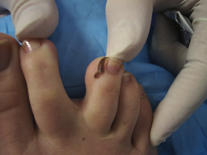
During the course of a tightly scheduled office day, a 30-year-old female presents with a painful paronychia involving the lateral border of her right hallux. The painful nail border is acutely inflamed. The doctor temporarily defers a definitive chemical matrixectomy and opts to perform a “slant-back” procedure to remove the offending nail border. The doctor adducts the patient’s foot ever so slightly to access the problematic portion of the affected nail unit more easily. While doing so, the clinician notices a tan/brown, slightly elevated papule inferior to the lateral malleolus. The lesion is not particularly large (6 mm) and is of uniform color. However, it exhibits moderate asymmetry of shape. The patient says the lesion had been present her “entire life.” The physician removes the offending nail border and the patient goes home believing that her pedal problems are now resolved. This general scenario is not uncommon. The patient may be a 75-year-old man and the skin lesion may be a small scaly plaque, but the net result is often the same: an early evolving malignancy goes undiagnosed. Incidentally, the aforementioned 30-year-old woman was a real patient but, in this instance, the podiatric clinician insisted on performing a biopsy on the atypical lesion. The biopsy disclosed an early evolving melanoma.
Factors That Can Lead To Incorrect Diagnosis
Readers might question how this could be possible. The patient (a young and competent historian) clearly stated the lesion had been there her entire life. This question leads us to the first important point: some melanomas arise in association with previously benign melanocytic nevi. Clinically atypical lesions must be subject to a histopathologic investigation. I have seen “well-intentioned” and competent clinicians who have been led astray and fell victim to “well-intentioned” and competent historians. This scenario is particularly common when dealing with geriatric patient populations. The simple fact is that melanoma may arise within longstanding, benign pigmented lesions on the skin, the skin of the foot not excluded. The history of longevity should not necessarily preclude obtaining a biopsy if one notes clinical atypia. A second point is that cutaneous malignancies are not “rare” in the skin of the foot as many of us were led to believe during our didactic years. Although malignancies related to sun exposure are less common on the foot, they may occur in this location. Melanomas arise in the skin of the lower extremity in 9 percent of cases within the Caucasian population and in half of all cases among African-Americans.1 Overall, as much as 15 percent of all melanoma is of the acral lentiginous type.2 Unfortunately, when melanomas arise on the skin of the foot, they do carry a poor prognosis largely due to delays in diagnosis.3 If a podiatric physician does not routinely diagnose skin cancer in his or her patient population, it is probably not that he or she hasn’t seen cancer. Rather, it is much more likely due to missed diagnoses. In some cases, we are “bailed out” by other medical professionals who are in a position to make the diagnosis in our stead. However, in many instances, we are not. We must bear in mind that the skin of the foot is foremost the responsibility of the podiatrist and much of the medical community has grown to depend on podiatrists in this regard. Unfortunately, it is all too common in mainstream medicine for patients to receive a “full body” dermatological exam with their socks on. As a result, I have seen numerous cases of melanoma and carcinoma involving the skin of the foot that went undiagnosed by dermatologists or general practitioners only to be picked up by an astute podiatrist. This will progress to become the norm as the podiatric profession continues to incorporate more and more of the techniques that have traditionally been limited to dermatological practices. Currently, dermatologically savvy DPMs stand apart in our profession simply because they recognize what is clinically atypical.
Atypical Lesions: What Should You Look For?
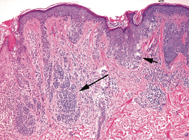
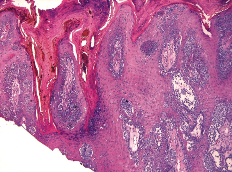
What should be considered clinically atypical when assessing pigmented lesions of the skin? As a general rule, we in podiatry too often fall into an unfortunate diagnostic trap. We fail to take note of dermatological tumors and other abnormalities until they resemble the classic textbook picture. This becomes particularly problematic on the skin of the foot as many common tumors will never have a classic appearance. For instance, due to the thick stratum corneum on the sole and the shearing forces that are imposed upon the skin of this location, nodular melanomas of the plantar surface are just as likely to resemble a foreign body as they are a classic melanoma. I have seen this several times. Alternatively, amelanotic lesions may be clinically identical to pyogenic granulomas. When they involve the nail bed, they may be indistinguishable from a subungual hematoma. Some clinicians are surprised to learn melanoma may also be profoundly verrucous in its configuration when arising in the skin of the sole. We recently reviewed a case in which a dermatologist elected not to biopsy what was believed to be a hemorrhagic verruca. Noting the clinical atypia (spontaneous hemorrhage, verruca arising in a middle-aged patient), an attentive podiatrist subsequently performed a biopsy on what was actually verrucous melanoma. The bottom line is if we calibrate our index of suspicion with what we see in the textbooks, then we will uniformly miss unconventional or more subtle presentations of melanomas. This is where the train wrecks occur and it is our patients who are inevitably picked from the wreckage. The same is true for squamous cell carcinoma and basal cell carcinoma. Thankfully, in most cases, the consequences for missing carcinomas on the pedal skin are usually not so monumental. Due to the lack of conventionality associated with melanoma of the lower extremity, it is not prudent to solely and strictly apply the criteria that define clinical atypia when assessing pigmented lesions elsewhere on the body. That being said, the traditionally applied criteria remain of great import. Foremost among the traditional indicators of atypia is asymmetry. In regard to lesional asymmetry, we may be referring to either the overall shape of the lesion in question or the distribution of its pigment. When pigmented lesions are bisected in any perpendicular plane, if the two resultant halves are not mirror images of each other, this is clinically atypical and should prompt us to examine the lesion more closely or perform an investigative biopsy. In some instances, the lesional asymmetry is a function of eccentric elevations or papules within an otherwise evenly contoured area of pigmentation. Some pigmented lesions may be quite symmetrical in shape but may have variation or asymmetry in color. The “red, white and blue” color spectrum that is written about in many textbooks is an extreme that is often not present, even in association with bona fide malignant melanoma. Clinicians should keep in mind that variation and/or asymmetry of color is usually much more subtle than this. Atypical-appearing nevi and melanoma are far more likely to exhibit areas of slight paleness or slight hyperpigmentation that is well within the spectrum of a tan-brown color. The third clinical feature that should prompt one to consider an investigational biopsy is irregular borders. As a general rule, benign melanocytic nevi possess regular crisp margins. Indistinct or “fuzzy” borders are, in and of themselves, atypical findings. Similarly, lesions with scalloped peripheral contours should not be considered routine. These findings may be subtle and far removed from the classic textbook photographs with which we are all familiar. If one cannot definitively trace a pigmented lesion with an ultra fine-tipped pen, one should promptly pursue further clinical and/or histopathologic investigation. A large diameter is the final traditionally described criterion that is an indicator of atypia in the realm of pigmented lesions of the skin. One must closely evaluate pigmented lesions that measure 6 mm in diameter or larger for additional atypical features, and consider performing an investigative biopsy. Studies have shown that when pigmented lesions of the palms and soles measure 7 mm in diameter or greater, the chance of melanoma is significant enough to make an investigational biopsy mandatory.4 Of course, there are other features that should warrant concern on the part of the podiatric clinician. When lesions show a tendency toward spontaneous hemorrhage or ulceration, clinicians should assume malignancy until proven otherwise. Simply stated, benign lesions do not, as a general rule, spontaneously ulcerate or bleed. When one encounters areas of foreign body-like pigment without a clear history of trauma, physicians should maintain a high degree of suspicion. When podiatric physicians are confronted with verrucous lesions arising in odd locations or exhibiting areas of altered pigmentation in people who are middle-aged or older, they should have an abundance of caution. Additionally, one should not dismiss pyogenic granuloma-like lesions that arise in older patient populations or show recalcitrance to treatment. In many instances, when such clinically atypical features are apparent, a diagnostic biopsy or conservative excision is in order. Finally, there is no replacement for the attention and clinical experience of the podiatric physician. One must investigate lesions that arise de novo or change in the appearance. Since changes may be subtle, clinical photographs may be quite useful, particularly when a biopsy is deferred. Such images provide a baseline that one can compare the lesion to on future dates. Do not think of pictures as a replacement for biopsy when evaluating pigmented lesions. If there is frank clinical atypia, pursue histopathologic investigation of pigmented lesions without delay. If a biopsy is deferred because clinicians elect to monitor the lesion for growth or changes, they should ensure appropriate follow-up.
Pertinent Pointers On Performing Biopsies
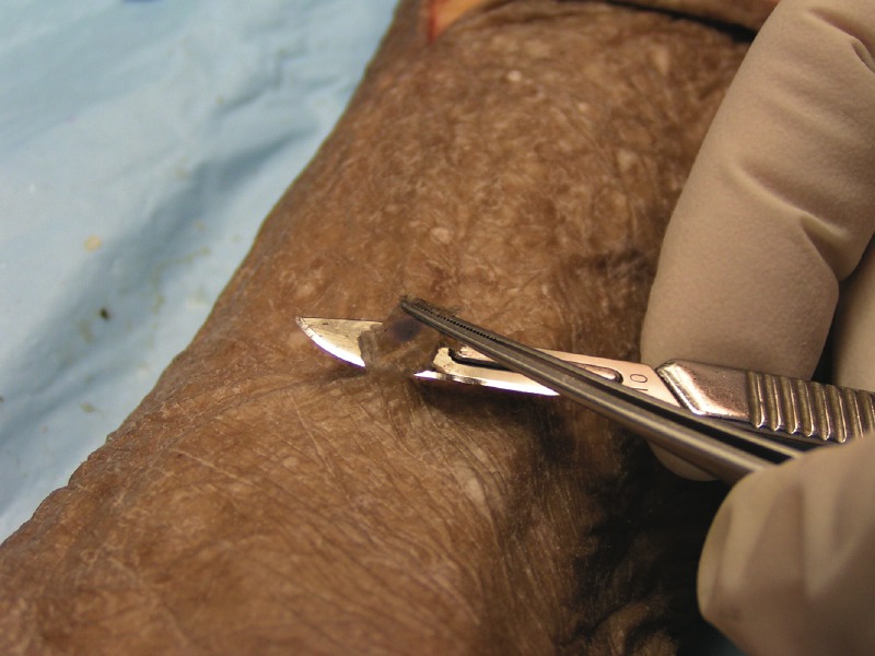
What is the biopsy technique of choice when sampling pigmented lesions? When assessing atypical pigmented lesions of the pedal skin, the single most important principle is making the correct diagnosis. From a clinical perspective, this means noting atypical features and subsequently performing an investigational biopsy. Ideally, the biopsy should sufficiently sample the lesion in question and allow for an accurate histopathologic interpretation. Although some biopsies may be of lesser quality than others, the single worst mistake a clinician can make is not the procurement of a subpar sample, it is failing to investigate an atypical pigmented lesion at all. However, quality is important as poor samples may mislead even the most skilled dermatopathologist. The goal of cutaneous biopsies is to guide definitive therapy. Sometimes biopsies performed on intermediate-sized or large lesions are intended only to obtain a representative sample. Alternatively, clinicians may often completely remove small pigmented lesions with a simple shave biopsy or ellipse, thus eliminating the need for a future excision if such an excision would have been mandated due to its histopathologic features. It might surprise some that even when complete removal is desired, only about 5 percent of dermatologists in the American Academy of Dermatology perform elliptical excision.5 The overwhelming majority of these cases are managed with either a standard shave biopsy or saucerization. The advantages of shave techniques include their ease and rapidity to perform, the lack of suture closure, minimal scarring and excellent patient tolerance. In addition, these techniques provide thorough sampling of the dermal-epidermal junction. Sampling this skin layer is of paramount importance because this region must be scrutinized by the dermatopathologist to discriminate effectively between melanomas and benign melanocytic nevi. Where an average punch biopsy samples 3.5 mm of the dermal-epidermal junction, shave biopsies often provide specimens of two to three times greater diameter. When punch biopsies are the chosen technique, especially when sampling much larger lesions, it is imperative to effectively communicate to the dermatopathologist the size of the lesion you are sampling. In difficult cases, such clinical findings may influence the diagnosis or the recommendations for future therapy. Many clinicians have asked me, “If it is atypical, why not just cut it out with an ellipse?” The answer is that this is a perfectly reasonable approach. The problem I have seen that when clinicians limit their biopsies to 3:1 ellipses with suture closure, they simply do not perform biopsies on the more subtle lesions. Since clinicians tend to under-biopsy, the less obvious malignancies go unsampled and thus undiagnosed. Many of us would be better served by learning from the dermatology profession and keeping our biopsies quick and simple.
What You Should Know About Shave Biopsies
Shave biopsies are excellent techniques when sampling superficially located tumors such as small to intermediate-sized pigmented lesions and exophytic (outward growing) tumors that form papules or nodules. These procedures may also be of utility when biopsying verrucous lesions or flat non-pigmented skin abnormalities such as superficial basal carcinoma, squamous cell carcinoma in-situ, or actinic keratosis (early-evolving squamous cell carcinoma in-situ). As with all biopsies of skin, the secret to performing standard shaves and saucerizations is to obtain the necessary tissue samples while keeping the procedures simple. Prefabricated “biopsy packs” are cumbersome to prepare and have unnecessary expense. Similarly, surgical drapes and timely pre-biopsy preps are not necessary in most cases. One would begin a shave biopsy by prepping the biopsy site with an alcohol wipe. Use lidocaine with epinephrine to anesthetize the biopsy site by infiltrating the dermis deep to, and immediately adjacent to, the biopsy site. For standard shave biopsies, use a scalpel equipped with a #10 or #15 blade to remove the necessary tissue. Hold the scalpel blade at a 15 to 30 degree angle to the skin surface. Tangentially score the epidermal surface and superficial dermis adjacent to the site to be sampled. This creates an epidermal “lip.” One may grasp this loose lip with forceps, allowing the scalpel to undermine the desired specimen with a mid-dermal plane of section. The surgeon should at least focally see glistening white dermal collagen at the base of the biopsy site. Once you remove the desired tissue, apply pressure to the biopsy site in conjunction with the chemocautery agent of choice. Saucerization technique differs from the standard shave only in that one performs this with a single sided razor blade or other blades commercially available for this purpose. Bow the blade between the thumb and middle finger using the index finger to stabilize it. Similar to the standard shave technique, the blade should enter the skin adjacent to the lesion in question. Then “scoop out” the desired sample, taking care to maintain a superficial or mid-dermal plane of section. Then apply hemostasis to the biopsy site and bandage it in the usual fashion.
Final Notes
Once one has taken the biopsy, there is an important decision of where to send the specimen for histopathologic evaluation. I believe clinicians should entrust their tissues to a person and not a lab. Ideally, clinicians should know the skills and qualifications of the person whose name is found at the bottom of his or her pathology reports. In addition, it is imperative that clinicians realize that all pathologists are not trained in the same manner. Board certification in anatomic pathology does not ensure competency when dealing with difficult melanocytic neoplasms. The same may be said for subtle dermatoses. Only board certification in dermatopathology will allow clinicians to be confident that their tissues are being reviewed by an experienced dermatopathologist. In the same vein, there is no advantage to using pathology labs that have dermatopathologists on staff unless it is he or she who is issuing the patient’s report. As roughly 25 percent of all the litigation involving melanoma is precipitated by a pathologist’s error, clinicians should demand that exclusively board-certified dermatopathologists review their skin biopsies.6 Finally, there is no substitute for communication. Dermatopathologists should be accessible and clinicians should feel comfortable calling them with questions or concerns. This type of interaction is irreplaceable when aspiring to offer the very best in medical care to the patients for whom we care. Dr. Bakotic is the Director of the Institute for Podiatric Pathology in Pompano Beach, Fla. He is also a Diplomate of the American Board of Pathology. References 1. Cress RD, Holly EA. Incidence of cutaneous melanoma among non-Hispanic whites, Hispanics, Asians, and Blacks: an analysis of California cancer registry data, 1988-1993. Cancer Causes Control. 8(2):246-252, 1997. 2. Weedon D. (ed) Lentigines, nevi, and melanoma In Skin Pathology. pp. 823. Churchill Livingstone: Edinburgh, 2002. 3. Cascinelli N, Zurrida S, Galimberti V, Bartoli C, Bufalino R, Del Prato I, Mascheroni L, Testori A, Clemente C. Acral lentiginous melanoma. A histological type without prognostic significance. J Dermatol Surg Oncol. 20(12):817-22, 1994. 4. Saida T, Ishihara Y, Tokuda Y. Effective detection of plantar malignant melanoma. Int J Dermatol. 32(10):722-725, 1993. 5. Tripp JM, Kopf AW, Marghoob AA, Bart RS. Management of dysplastic nevi: a survey of fellows of the American Academy of Dermatology. J Am Acad Dermatol. 46(5):674-82, 2002. 6. Jackson R. Malignant melanoma: a review of 75 malpractice cases. Int J Dermatol. 36(7):497-498, 1997. For related articles, check out the archives at www.podiatrytoday.com.
References:
**sub**CE Exam #129**endsub** Choose the single best response to each question listed below: 1. Melanoma occurs in the lower extremity in what percentage of melanoma cases in African-Americans? a) 9 percent b) 15 percent c) 20 percent d) 50 percent 2. Amelanotic lesions may resemble … a) pyogenic granulomas b) hemorrhagic verruca c) squamous cell carcinoma d) all of the above 3. Foremost among the traditional indicators of atypical pigmented lesions is … a) asymmetry b) regular crisp margins c) lesions smaller than 3 mm in diameter d) none of the above 4. One should closely scrutinize pigmented lesions that are _ mm or larger for atypical features and consider an investigative biopsy. a) 6 b) 7 c) 8 d) 9 5. Fifteen percent of all melanoma is … a) melanocytic b) atypical c) of the acral lentiginous type d) verrucous 6. As a general rule, benign melanocytic nevi possess … a) indistinct borders b) lesional asymmetry c) regular crisp margins d) none of the above 7. Which of the following is an advantage of performing a shave biopsy? a) Minimal scarring b) Lack of suture closure c) Easy and quick to perform d) All of the above 8. When amelanotic lesions involve the nail bed, they most often resemble … a) subungual hematoma b) actinic keratosis c) pigmented basal cell carcinoma d) benign melanocytic nevi 9. One is more likely to see which color change in relation to malignant melanoma? a) Areas of brown hyperpigmentation b) Areas of blue hyperpigmentation c) Areas of red hyperpigmentation d) Areas of near-white hyperpigmentation Instructions for Submitting Exams Fill out the enclosed card that appears on the following page or fax the form to NACCME at (610) 560-0502. Within 60 days, you will be advised that you have passed or failed the exam. A score of 70 percent or above will comprise a passing grade. A certificate will be awarded to participants who successfully complete the exam. Responses will be accepted up to 12 months from the publication date.


