

ADVERTISEMENT
A Closer Look At Bone Graft Substitutes
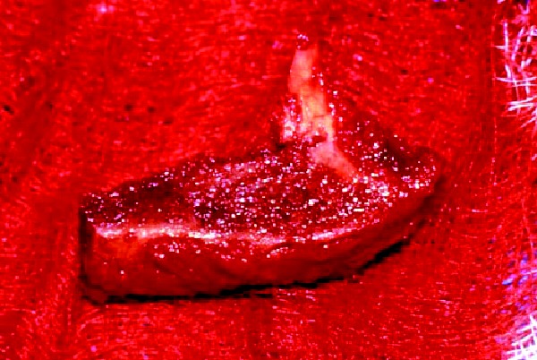
There are many instances when it is appropriate for the foot and ankle surgeon to use bone grafts in order to enhance a patient’s clinical outcome. When it comes to reconstructive osteotomies for angular realignment, arthrodeses and the repair of nonunions and cystic bone lesions, these are often best performed with procedures that take advantage of the many desirable features of bone grafts and, more recently, bone graft substitutes. Before discussing the details of bone graft substitutes, it is helpful to review the bone graft options that are available to the surgeon. These options include: cancellous and/or cortical bone, allogeneic (typically freeze-dried) cadaver bone and autogenous bone. Cancellous bone offers a porous, osteoconductive environment and ease of incorporation. However, it conveys limited structural strength and ability to retain fixation devices. Cortical bone, on the other hand, offers tremendous structural integrity, but it is slow to incorporate in comparison to cancellous bone. Allogenic bone implants offer ease of use, ready availability and provide a wide range of configurations that include cortical and cancellous bone in various proportions. However, despite rigorous measures related to the procurement and preparation of allogeneic tissue products, there remains a very small but real risk of disease transmission and hypersensitivity reaction.1,2 The autogenous corticocancellous bone graft, which is typically harvested from the iliac crest, remains the gold standard for comparison of all other grafting materials. In reconstructive foot and ankle surgery, the calcaneus offers a reliable source of autogenous bone graft material. Unfortunately, however, autogenous sources of corticocancellous bone are limited and there is distinct morbidity associated with harvesting autogenous bone. Banwart et al., performed a meta-analysis of the related literature and observed that use of autogenous iliac crest bone grafts was associated with a 25 to 45 percent complication rate. They also noted that 40 percent of patients related harvest site pain up to five years postoperatively.3 For these reasons, the emergence of bone graft substitutes for enhancement or augmentation of bone grafting procedures, as well as the techniques of bone callus distraction, have proven to be of great service in recent years.
Understanding The Physiological Processes Of Bone Healing
In order to appreciate the useful features of bone graft substitutes, it is important to review some of the physiological processes underpinning bone healing in the presence of a bone graft. Bone graft incorporation occurs via a process referred to as “creeping substitution,” whereby the graft material is actually replaced by new bone formation. This desirable and complex event depends upon three underlying processes: osteoconduction, osteoinduction and osteogenesis. Osteoconduction refers to the process in which osteoprogenitor cells and vascular elements migrate to and adhere along a suitable porous, inorganic scaffold. Osteoinduction refers to the process in which undifferentiated osteoprogenitor stem cells are recruited and migrate to the site of bone formation. Once these stem cells are present along the porous substrate, they differentiate into bone producing cells such as chondroblasts (enchondral bone formation) and osteoblasts. Osteogenesis refers to the process in which bone producing cells, either presenting directly (as osteocytes, osteoblasts or chondroblasts) or indirectly from previously undifferentiated stem cells, create new bone by laying down osteoid or via enchondral calcification of cartilage. (See “A Guide To Bone Regeneration And Graft Healing” below.) With these processes in mind, we will now explore many of the bone graft substitutes available to foot and ankle surgeons. Graft substitutes entail a variety of products, ranging from allogeneic to autogenous materials. One may use these products separately or in combination with other substitute materials or with allogeneic or autogenous bone grafts. Indeed, investigation into the results of combining osteoconductive and osteoinductive materials has shown healing rates that are compatible with those observed with autogenous bone grafts.4,5 When one combines osteoconductive substitute materials with traditional bone graft materials, surgeons may think of these substitute materials as “bone graft expanders.” When one combines inductive and conductive materials, surgeons may think of these materials as “bone graft enhancers.” The combination of mesenchymal stem cells and/or living osteogenic cells with osteoconductive and osteoinductive materials creates a true bone graft substitute.
What You Should Know About Osteoconductive Materials
Osteoconductive materials include the inorganic bioceramics, calcium phosphate and calcium sulfate as well as the hydroxyapatites. These materials are available in many forms, including powders, granules, blocks and morsels. Calcium phosphate has been used in bone graft surgery for over 25 years.6 The range of pore sizes (1-1,000 microns) in calcium phosphate closely resembles that of human cancellous bone. In essence, calcium phosphate mimics the osteoconductive pores of human cancellous bone. Moreover, calcium phosphate is rapidly (10 to 12 weeks) reabsorbed in vivo. Similar to other synthetic bioceramics, calcium phosphate is sterile and biocompatible. Therefore, it is completely safe relative to disease transmission and immunogenicity. Typical uses of calcium phosphate include back-filling procedures following evacuation of bone cysts, as well as filling the void created when one has harvested autogenous cancellous bone. Calcium phosphate osteoconductors include Skelite™ (EBI) and Vitoss™ (Orthovita). Calcium sulfate also provides a useful osteoconductive substrate that differs from calcium phosphate in that it is considerably more dense after curing. When properly used, the porous conductive properties of calcium sulfate are very similar to those of calcium phosphate. However, after an initial pliable period, calcium sulfate hardens to a dense filler that one may use to fill defects and support purchase by fixation devices. Like calcium phosphate, calcium sulfate reabsorbs completely and rapidly.7 One may also add antibiotics to calcium sulfate and use the combination to pack debrided bone defects when managing osteomyelitis. Calcium sulfate osteoconductors include Osteoset™ (Wright Medical) as well as AlloMatrix™ and miniMIIG™ (Wright Medical), which are combined with demineralized bone matrix (DBM). Hydroxyapatite, either as natural coralline hydroxyapatite or synthetic calcium carbonate, also provides an inorganic osteoconductive porous scaffold suitable for back-filling procedures.8 Hydroxyapatite is inorganic and non-immunogenic. It provides minimal structural strength, undergoes relatively slow biodegradation, and one can observe this radiographically even at 18 months after implantation. Hydroxyapatite osteoconductors include Interpore 200 (Interpore International) and Pro Osteon 500™. The combination of tricalcium phosphate (35 percent) and hydroxyapatite (65 percent) in a nonstructural allogeneic collagen matrix (Collagraft™, Zimmer) provides a bone graft expander that is readily available and useful for placing within or about a large arthrodesis interface or as a back-filling osteoconductor.
Assessing The Potential Of Osteoinductive Materials
The mainstay of osteoinduction therapy is DBM. DBM is derived from demineralized allogeneic cadaver bone or from recombinant, human-cloned bone matrix.9 There can be variability in the bioactivity of DBM from batch to batch, and the various proprietary processes used by different manufacturers are important in this regard.10 In particular, the debate persists over whether terminal gamma (high energy) versus electron beam (low energy) irradiation diminishes the bioactivity of DBM. This debate will probably continue until a head-to-head, randomized controlled clinical trial or a large observational study compares the results of bone graft healing using the two different forms of DBM. Since DBM is allogeneic, there is a very small potential for transmission of infection and the possibility of symptomatic immunogenicity. DBMs contain bone growth factors that signal and modulate the processes of bone healing and graft incorporation. These include the growth factors BMP 2 and 7 that regulate mesenchymal stem cells and osteoblasts, and also stimulate angiogenesis. BMPs are available in many forms, including powder, gel, putty and flexible sheets of demineralized bone collagen fibers impregnated with BMPs. One may also combine DBM with bioceramic and/or hydroxyapatite substrates to expand and enhance graft healing. Surgeons may use commercially available osteoinductive products alone or in combination with osteoconductive substrates. These osteoinductive products include: Grafton™ (Osteotech), DBX™ (Synthes), Osteofil™ (Regeneration Technologies, Inc.), OsteoStim (EBI) and the aforementioned Allomatrix. One may employ platelet gels to concentrate platelets and, hence, growth factors that influence bone healing (as well as healing in other tissues). Concentrated platelets can be useful for osteoinduction, clot stabilization and chemotaxis (signaling undifferentiated stem cells to the bone graft). Platelet rich plasma, which is obtained by spinning autologous blood from venipuncture in a centrifuge, provides a variety of inductors that play a part in bone graft healing. These include: platelet-derived growth factor; transforming growth factor beta, which is responsible for transforming undifferentiated stem cells into osteogenic cells; and vascular endothelial growth factor. Commercially available autologous platelet separator systems include Symphony™ (DePuy) and Magellan™ (Medtronic).
Do Osteogenic Materials Have A Place In Bone Healing?
Osteogenesis can only be conveyed to the bone graft via living stem cells, osteoblasts or chondroblasts. Currently, the most readily available source of such cells is autogenous aspiration from medullary bone. There are higher concentrations of undifferentiated stem cells in younger individuals (with the highest concentrations being present in children) and in bones more centrally located in the axial skeleton.11,12 Therefore, a bone marrow aspiration of the ilium will, on average, contain more undifferentiated stem cells than the femur, which will contain more than the distal tibia or calcaneus. For this reason, a more proximal marrow aspiration is preferable to a distal aspiration. Depending upon the direction of future research efforts, perhaps cloned human stem cells will become the main source of osteogenic capacity for bone grafts. Currently, autogenous aspiration of stem cells is the most readily available source. Although one may employ a generic bone marrow aspiration needle to harvest medullary stem cells, commercially available systems provide enhanced features that coordinate the aspiration process with bone grafting. These systems include Imbibe Bone Marrow Aspiration Syringe™ (Orthovita) and I/C Graft Chambers™ (DePuy).
Final Notes
Although bone graft substitutes offer materials that surgeons can use to enhance bone graft healing, they do not replace or obviate the need for strict adherence to the principles of osteosynthesis and a variety of other interventions that are useful in managing bone healing. These measures include but are not limited to: • electrical (or sonic) bone growth stimulation; • bisphosphonate therapy for Charcot or osteoporotic individuals; • appropriate nutritional supplementation (particularly with respect to zinc and calcium); • control of anemia; • discontinuation of tobacco smoking; • assurance of satisfactory extremity vascularity; • avoidance of the disruptive forces of weightbearing; and • the proper balance of immobilization and physical rehabilitation. It is also very important for patients to understand the importance of compliance with the therapeutic regimen. In summary, bone graft substitutes offer foot and ankle surgeons materials that meet the desirable attributes of a bone graft while reducing the risk of donor site morbidity. These materials convey the properties of osteoconduction, osteoinduction, and osteogenesis. One would typically employ these materials in combination with autogenous and/or allogeneic bone graft materials, or other substitute materials. An area of active biomedical research, bone graft substitutes provide surgeons with a useful method for improving the results of bone graft healing without the need, in many cases, for harvesting large, autogenous, corticocancellous bone grafts. Dr. Malay is a Fellow of the American College of Foot and Ankle Surgeons, and is in private practice at the Ankle and Foot Medical Centers of the Delaware Valley. He is the Director of Podiatric Research at the University of Pennsylvania Medical Center-Presbyterian, and is a Fellow of the Center for Clinical Epidemiology and Biostatistics at the University of Pennsylvania. Dr. Malay is also a faculty member of the Podiatry Institute.
References:
1. Bauer TW, Smith ST. Bioactive materials in orthopaedic surgery: overview and regulatory considerations. Clin Orthop. 2002 Feb;(395):11-22.
2. Anonymous, EEC regulatory document. Note for guidance. Validation of virus removal and inactivation procedures. Committee for Proprietary Medicinal Products: Ad Hoc Working Party on Biotechnology/Pharmacy and Working Party on Safety Medicines. Biologicals. 1991 Jul;19(3):247-51.
3. Banwart JC, Asher MA, Hassanein RS. Iliac crest bone graft harvest donor site morbidity. A statistical evaluation. Spine. 1995 May 1;20(9):1055-60.
4. Skoff HD. Bone marrow/allograft component therapy. A clinical trial. Am J Orthop. 1995 Jan;24(1):40-7.
5. Gazdag AR, Lane JM, Glaser D, Forster RA. Alternatives to Autogenous Bone Graft: Efficacy and Indications. J Am Acad Orthop Surg. 1995 Jan;3(1):1-8.
6. Cavagna R, Daculsi G, Bouler JM. Macroporous calcium phosphate ceramic: a prospective study of 106 cases in lumbar spinal fusion. J Long Term Eff Med Implants. 1999;9(4):403-12.
7. Alexander DI, Manson NA, Mitchell MJ. Efficacy of calcium sulfate plus decompression bone in lumbar and lumbosacral spinal fusion: preliminary results in 40 patients. Can J Surg. 2001 Aug;44(4):262-6.
8. Demers C, Hamdy CR, Corsi K, Chellat F, Tabrizian M, Yahia L. Natural coral exoskeleton as a bone graft substitute: a review. Biomed Mater Eng. 2002;12(1):15-35.
9. Martin GJ Jr, New formulations of demineralized bone matrix as a more effective graft alternative in experimental posterolateral lumbar spine arthrodesis. Spine. 1999 Apr 1;24(7):637-45.
10. Glowacki J, Mulliken JB. Demineralized bone implants. Clin Plast Surg. 1985 Apr;12(2):233-41.
11. Muschler GF, Boehm C, Easley K. Aspiration to obtain osteoblast progenitor cells from human bone marrow: the influence of aspiration volume. J Bone Joint Surg Am. 1997 Nov;79(11):1699-709. Erratum in: J Bone Joint Surg Am 1998 Feb;80(2):302.
12. Muschler GF, Nitto H, Boehm C, Easley KA. Age- and gender-related changes in the cellularity of human bone marrow and the prevalence of osteoblastic progenitors. J Orthop Res. 2001 Jan;19(1):117-25.


