

ADVERTISEMENT
News and Trends
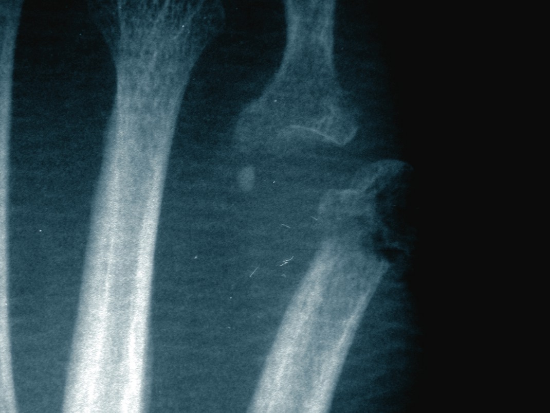
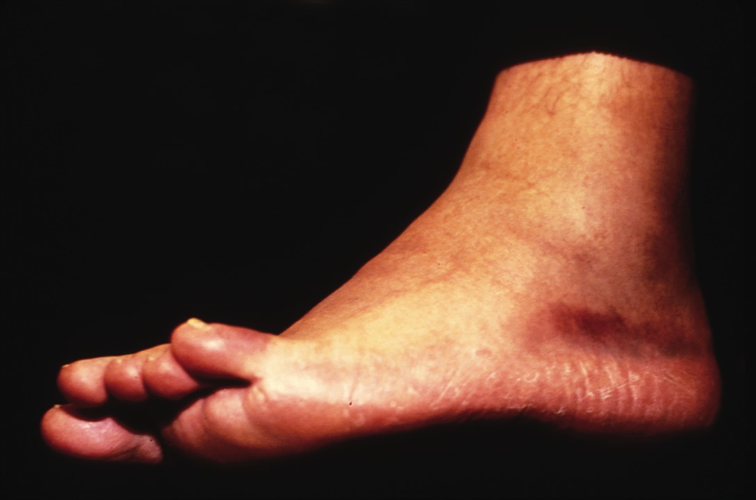
How Effective Is The PTB Test In Diagnosing Osteomyelitis?
There has been some recent debate within the profession about the effectiveness of the probe-to-bone (PTB) test in diagnosing osteomyelitis. A new study in Diabetes Care has found that the PTB test has a relatively low positive predictive value when it is utilized for diabetic patients with foot wounds. The two-year study tracked 1,666 patients with diabetes who underwent regular foot exams and were instructed to come to the clinic if they developed signs of lower-extremity complications. For those with infections, researchers compared PTB test results to a culture of infected bone. Study authors said the PTB test was positive if the bone or joint were palpable and they defined osteomyelitis as a positive bone culture. Over 27.2 months, 247 patients developed foot wounds and 151 patients had 199 foot infections, according to the study. Researchers found osteomyelitis in 30 patients or 12 percent of patients with foot wounds and 20 percent of patients with foot infections. For all diabetic foot wounds, the study says the PTB test was “highly sensitive” (0.87) and specific (0.91). The positive predictive value was 0.57 but the negative predictive value was 0.98. Investigators concluded that while the PTB test has a low predictive value for those with diabetic foot ulcers, a negative PTB test may exclude the diagnosis of osteomyelitis. Study co-author Benjamin Lipsky, MD, says one should conduct the PTB test as part of a thorough evaluation of any open foot wound. As one probes to find the ulcer depth and searches for any foreign bodies, he notes the ease of checking for the presence of palpable bone. Study co-author Lawrence Lavery, DPM, MPH, says although another simple bedside test would be effective, there is nothing in the pipeline. In many cases, he says diagnosing osteomyelitis comes down to clinical judgment and clinical correlation is “an essential component.” He notes that testing is one aspect of the process and labs, imaging studies, patient characteristics and the wound all contribute to the diagnosis of osteomyelitis.
When Are Additional Diagnostic Tests Needed?
As Dr. Lavery notes, bone biopsy is still the gold standard for osteomyelitis diagnosis and MRI or bone scans must meet or compare against the bone biopsy standard. “The risk is that osteomyelitis will often be over-diagnosed with imaging techniques. Previous surgery, Charcot arthropathy and trauma can all show false positives,” says Dr. Lavery, a Professor in the Department of Surgery at Texas A&M Health Science Center College of Medicine. If a DPM is assessing for osteomyelitis in the office, study co-author David G. Armstrong, DPM, PhD, advises considering other tests, such as serial radiography, MRI or bone biopsy, along with the PTB, depending on the circumstance. Dr. Armstrong notes that the majority of the time, osteomyelitis by itself is not an acute emergency and infection control, along with biopsy, MRI or serial radiographs (depending on the circumstance), will “win the day.” Often, one will make a presumptive diagnosis long before securing an expensive test, says Dr. Armstrong, a Professor of Surgery, Chair of Research and Associate Dean at the William M. Scholl College of Podiatric Medicine at Rosalind Franklin University of Medicine. “The clinician must weigh the expense of failing to make the right diagnosis, treating with the wrong antibiotic or wrong duration, or encouraging antibiotic resistance against the cost of any imaging studies,” says Dr. Lipsky, a Professor of Medicine at the University of Washington School of Medicine. For almost all patients with a diabetic foot ulcer, he feels plain X-rays are useful. However, when one has a reasonably high suspicion of osteomyelitis, perhaps a greater than 30 percent likelihood, and plain X-rays are not helpful, Dr. Lipsky says it is appropriate to consider more expensive imaging studies. In many instances, he says a bone biopsy is the best way both to definitively diagnose osteomyelitis and to determine the etiology pathogen and their antibiotic susceptibilities.
Addressing Common Misperceptions
In regard to the PTB test, Dr. Lavery says there is a tendency to generalize the findings from the 1995 Grayson study, in the Journal of the American Medical Association, which found a “very high prevalence of bone infections” with the PTB test. However, he emphasizes that the Grayson study assessed patients hospitalized for severe infection. Dr. Lavery says there has been a tendency to apply those PTB findings to a broader clinical population than the Grayson study included. “The recent Diabetes Care study did not come here to bury the PTB test but rather to praise it in its appropriate context,” adds Dr. Armstrong. “The PTB test is highly valuable. However, the bottom line is that it is less valuable by itself in an outpatient setting, where fewer people have osteomyelitis.” What questions should be explored in further research on the PTB test? Dr. Lavery says researchers must determine what variability exists among examiners when applying the PTB test. Dr. Lipsky concurs, noting that the profession does not know how reproducible the results are when more than one person does the test. He says considering the inter-rater reliability can note the true reliability of a “positive” or “negative” result.
PODIATRY IN PRACTICE: VA System Has Excellent Care For Vets, Advantages For DPMs
By Brian McCurdy, Senior Editor As Jeffrey Robbins, DPM, notes, the Veterans Health Administration (VA) is “the benchmark in the care of many chronic diseases, including diabetes and amputation prevention.” He says the VA includes numerous practice models, such as hospital-based practice and community based practice in Community Based Outpatient Clinics (CBOCs). Dr. Robbins says most of the new VA staff are “highly trained” and provide surgical and podiatric care, which includes biomechanics. Dr. Robbins, the Director of Podiatry Service at the VA Central Office in Cleveland, delineates the three missions of the VA system: providing excellent patient care to improve veterans’ quality of life; providing excellent education to benefit the next generation of healthcare providers for the VA and nation; and providing excellent research to improve the delivery of healthcare. As he notes, all the VA’s facilities provide opportunities to offer excellent patient care and many facilities train students and residents as well as offer research opportunities. As Dr. Robbins notes, the VA is the largest managed care system in the world and has “arguably the finest computer system” with the Computerized Patient Record System (CPRS). The system permits immediate access to all patient information and he notes that the CPRS manages referrals and prescriptions. “Every examination, progress note, operative report, discharge summary, results of consults, medications, lab test results, imaging results, clinical reminder and demographic information is right there at your fingertips,” explains Dr. Robbins. Having various specialists within the same building enables those needing immediate attention to get either a “curbside consult” by a specialist or a referral to the emergency department for admission if necessary, points out Dr. Robbins. He says this ensures care is not delayed because of issues with travel, availability or access. “The single most important advantage to working in the VA is that our patient population are real life heroes,” adds Dr. Robbins. He emphasizes that they “placed themselves in a position where they either could have been, or were, in harm’s way to protect our way of life. Regardless of your politics, these people were there for us and now we are here for them.”
What Are The VA’s Challenges And Rewards?
Working for the federal government also includes some economic advantages, according to Dr. Robbins. Salaries are reasonable depending on geographic location and Dr. Robbins says the VA offers a “very good” benefits and retirement package. An additional benefit is that the VA podiatrists do not need to worry about paying for practice supplies, personnel, rent, equipment, utilities or managing a small business. Likewise, DPMs do not worry about third-party payers covering their work although Dr. Robbins says they do have to code properly since the system does bill third-party payers other than Medicare. Dr. Robbins notes that the VA’s biggest challenge is in passing a legislative initiative to move the podiatry service under the same hiring authority as medicine and dentistry so that podiatrists would qualify for the same pay structure so the system can still recruit “the best and brightest” into its ranks. As Dr. Robbins points out, the VA system includes “an impressive workforce” with lecturers, authors, educators and other lower extremity authorities. The patient load for the VA is also increasing along with the severity of the problems. Accordingly, Dr. Robbins says they are developing a basic foot care course for non-physician providers to help address the increasing number of patients seen in the VA system. “We are developing a basic foot course for non-physician providers to help provide basic foot care under our supervision so that our podiatrists are able to see the more complex conditions that present,” explains Dr. Robbins. Dr. Robbins says the VA is innovative and encourages contributions from its staff. “Like any system, it has its problems and there are a very small number of areas that are still not up to speed. For the most part, however, this is a system that is moving forward, setting the standard and is determined to improve the quality of life for America’s veterans,” emphasizes Dr. Robbins. For more of the “Podiatry In Practice” series, check out the January and February “News and Trends” sections.
Study: MRI Can Change Ankle Pain Diagnosis
By Brian McCurdy, Senior Editor Can the use of magnetic resonance imaging (MRI) provide valuable insight into ankle injuries? A recent study in the American Journal of Roentgenology says MRI can potentially change diagnoses in patients with ankle injuries and possibly lead to less invasive treatments. Researchers studied 91 patients who were referred from one orthopedic foot and ankle surgeon for ankle MRI. The study concluded that MRI findings changed the management plans for 35 percent of patients in the study. For 32 percent of patients for whom management plans changed, practitioners formulated a less invasive plan, according to the study. The surgeon from whom patients were referred believed that in 66 percent of cases overall, the understanding of the patient’s condition had either depended on the MRI or had been improved by the imaging. Babak Baravarian, DPM, uses MRI studies “a great deal” when treating ankle pain. He says in cases of instability, there are frequent secondary findings like synovitis, osteochondral lesions or peroneal tendon injuries. “With good readings, a great deal of information is gained from an MRI study,” says Dr. Baravarian, the Co-Director of the Foot and Ankle Institute of Santa Monica. “I think the info is essential prior to most surgical cases about the ankle.” Lawrence A. DiDomenico, DPM says as costs decrease, it is easier to use the technology from an insurance standpoint. Increasing MRI use is becoming a more common and accepted practice pattern for musculoskeletal physicians. In the ‘80s, he recalls there was not much literature on specific soft tissue pathologies such as plantar fascia ruptures or peroneal tendon injuries. Dr. DiDomenico, an Adjunct Professor with the Ohio College of Podiatric Medicine, says MRI has made these conditions easier to diagnose, resulting in a recent increase in the reports in the literature. He adds that DPMs now see these pathologies more frequently. “It gives a much more accurate diagnosis and therefore, more focused treatment options for patients,” he says. Dr. DiDomenico says a facility without a well-trained musculoskeletal radiologist and/or MRI technologist may generate a poor reading (i.e., a one- or two-sentence report). Alternatively, more experienced staff will provide a detailed one- or two-page report, according to Dr. DiDomenico, the Director of the Reconstructive Rearfoot and Ankle Surgical Fellowship with the Ankle and Foot Care Centers in Youngstown, Ohio.
Study Examines Acellular Regenerative Tissue Matrices
When a patient presents with a diabetic midfoot wound in combination with a midfoot Charcot deformity, podiatrists may face a potential complication of increased peak pressures and delayed wound healing. However, one may find success using an acellular regenerative tissue matrix to treat the condition, according to an abstract to be presented at the Symposium on Advanced Wound Care (SAWC) next month. For the abstract, researchers examined 10 patients with diabetes, recalcitrant wounds that had failed local wound care and a collapsed midfoot. After patients underwent posterior tibial tendon reconstruction, an acellular regenerative tissue matrix was utilized and the patients received continuous wound care for a year. After the year of follow-up, the abstract notes that all patients experienced full midfoot healing without wound recurrence and wound healing occurred at an average of three months. The midfoot alignment between the bisection of the talus and metatarsal remained almost parallel with no signs of collapse, and all patients experienced increased strength and stability of the posterior tibial tendon compared to the contralateral limb, according to the abstract. The author notes no complications from the procedure and says patients tolerated the post-op course well. How can acellular regenerative tissue matrix (ARTM) aid such patients? Abstract author Gerit Mulder, DPM, MS, says ARTM is a temporary tissue substitute that the host’s own cells eventually replace. As he notes, patients should experience an end result augmentation and potential joint resurfacing. ARTM’s contraindications are infection, underlying tissue necrosis and any sensitivity to the product, according to Dr. Mulder, an Associate Professor of Surgery and Orthopedics at the Department of Surgery/Division of Trauma at the University of California-San Diego (UCSD). The Symposium on Advanced Wound Care will be held from April 28 to May 1 at the Tampa Convention Center in Tampa, Fla. For more info, go to www.sawc.net.
In Brief
Scholl’s Center for Lower Extremity Ambulatory Research (CLEAR) at Rosalind Franklin University of Medicine and Science recently debuted a new CLEARcast series of podcasts. The CLEARcast will be released biweekly and its mission is reviewing important current literature in the field of lower extremity diabetes complications, according to CLEAR. The CLEARcast will interview authors of important papers, as well as others in the profession who are making pivotal contributions to foot-related research, clinical care and education, according to CLEAR. The next CLEARcast, now available, examines the Infectious Disease Society of America’s (IDSA) guidelines on diagnosing and treating diabetic foot infections, and features an interview with Warren Joseph, DPM. CLEAR says the CLEARcast is iPod/iTunes compatible and available free for download and subscription at www.diabetic-foot.net or the iTunes Music Store.


