

ADVERTISEMENT
Secrets To Treating Ankle Fractures In Athletes
More and more people are in the pursuit of becoming active and staying fit. More often than not, individuals tend to achieve this goal by participating in sporting activities. Whether they are participating in intramural or competitive activities, these athletes place a great demand on the ankles and feet. According to the National Collegiate Athletic Association Injury Surveillance System for 2000-2001, the ankle, knee and lower extremity were common sites of injury. The ankle joint is reportedly one of the most common sports-related injuries clinicians see in emergency rooms and private offices.1 Of these visits, ankle sprains make up 10 to 28 percent and ankle fractures occur 4 to 15 percent of the time.2-4 They account for approximately 3 to 5 million injuries occurring among competitive and recreational athletes in the United States, and cost nearly $1 billion annually in treatment.5 More often than not, most ankle injuries are sprains or strains and tend to heal with conservative therapy. When the athlete applies greater force on the ankle joint than the joint can resist, a fracture tends to occur. Although the reported incidence of ankle fractures in the athletic population is low, they tend to lead to deleterious results. Treatment of ankle injuries and fractures in the highly competitive athletes can be challenging because of the great demand placed on the ankle joint. Despite the tremendous amount of controversy, much attention has been focused on extrinsic and intrinsic risk factors for ankle injuries. Loss of playing time, decline in performance and the expectation to return to pre-injury status are areas in which athletes have great concern.2 Regardless of the competitiveness, the margin of error for accurate diagnosis and treatment of these patients is low. When these athletes present with an ankle fracture, one must assess and differentiate between closed versus open fractures and stable versus unstable fractures. Foot and ankle surgeons must also identify injuries to other soft tissue and/or osseous structures. After noting all of the above, clinicians must implement radiographic evaluation to determine the extent of the injury. Surgeons commonly use the Lauge-Hansen and Danis-Weber classification systems to describe ankle fractures, and employ the Berndt and Harty, and Salter-Harris classification systems to assess the degree of talar dome lesions and epiphyseal injuries respectively.5 Accordingly, let us review these classification systems, risk factors for ankle fractures, treatment options for ankle fractures and their respective outcomes. For this article, we will take a closer look at managing the athletic patient with a particular emphasis on rehabilitation and early weightbearing.
A Guide To Key Classification Systems
The Lauge-Hansen classification system is the most widely used system to assess ankle fractures.6 This system helps the physician visualize the mechanism of injury, the ability to reproduce the fracture, ascertain the anatomy involved and ability to reduce the fracture. (See “What You Should Know About The Lauge-Hansen Classification System” below.) This classification system was solely based on experimental cadaveric studies and clinical experiences with common fractures. The first term describes the position of the foot at the time of injury while the second term describes the direction of the pathologic force on the talus. The staging in this classification is as follows: supination-adduction, supination-eversion (supination–external rotation), pronation-abduction, pronation-eversion (pronation-external rotation).6 The Danis-Weber classification system is the second commonly utilized system among surgeons when it comes to identifying the degree of ankle fractures. However, the use of this system has increased in recent years due to the ease of classifying the fracture. The basis of this system is classified on the fracture pattern of the fibula and the integrity of the syndesmosis. This classification recognizes Type A, Type B and Type C fractures.6 With a Type A fracture, the fracture line is below the ankle joint level. These fractures account for 14.8 to 27 percent of all ankle fractures. Approximately 41 percent of ankle fractures are Type B fractures, in which the fracture line is at the level of the ankle joint. Type C fractures, which involve a fracture line below the level of the ankle joint, comprise about 20 percent of ankle fractures. The Berndt and Harty classification is the most commonly accepted system clinicians use for describing transchondral lesions. The defect often results from a shearing of the distal tibia and talar dome under compression that is often secondary to an ankle sprain or fracture. These types of injuries are difficult to recognize in acute phase due to the similar presentation of an ankle sprain. According to the Berndt and Harty classification, a stage 1 lesion is a trabecular compression fracture of subchondral bone; a stage 2 lesion is a partially detached osteochondral fragment; a stage 3 lesion involves a completely detached, non-displaced fragment; and a stage 4 lesion involves a detached and displaced fragment. In regard to assessing the degree of epiphyseal fractures in pediatric patients, the classification system proposed by Salter and Harris is the most widely accepted (see “A Closer Look At The Salter-Harris Classification System For Epiphyseal Fractures” below). The mean age for most physeal fractures is reportedly in the range of 1 to 16 years old with 10 years of age being the average.
Key Considerations With The Epidemiology
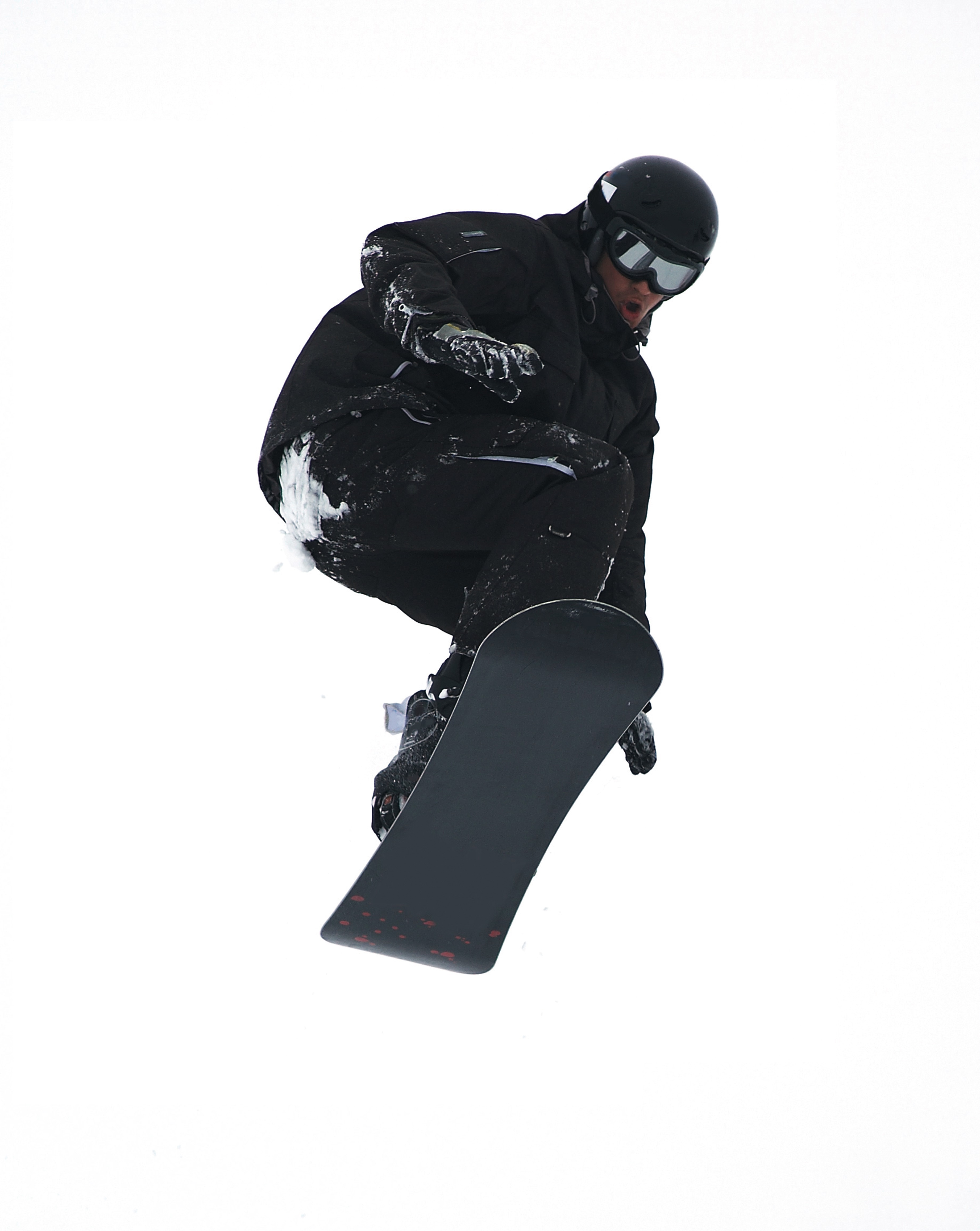
Chandler divided athletic activities into two groups based on the potential energy of injury: low-velocity sports and high-velocity sports.7 Low-velocity sports involve velocities less than 20 mph. These sports include track, swimming, tennis, volleyball, basketball, football and baseball, and rely on human propulsion for motion. Injuries in this category are usually limited to strains, sprains, simple non-displaced fibula fractures and some simple bony injuries. Therefore, it is best to handle the aforementioned injuries with conservative care. Ice, elevation, taping and aggressive rehabilitation program are often sufficient. If there is a simple bony injury, one can usually treat this with four to six weeks of casting or bracing. Severe fracture-dislocation problems are rare with these sports. High-velocity sports are events such as motor racing, downhill skiing, ice hockey, snowboarding, horse racing and skydiving. These sports involve velocities greater than 20 mph and require the use of machines and/or animals to produce acceleration exceeding human capabilities. Injuries in this category are generally displaced, comminuted or open fractures that involve other osseous structures and require surgical intervention followed by non-weightbearing.
Understanding The Impact Of Extrinsic Risk Factors
In the literature, there has been great emphasis on risk factors for ankle injuries. Barker says the most common risk factors are of great controversy.4 However, a review of the literature does appear to show some areas of common thought with extrinsic and intrinsic risk factors playing a major role.4,5 Examples of possible extrinsic risk factors include the level of competition, skill level, shoe type, use of ankle tape or bracing and the playing surface. Intrinsic risk factors are things such as age, ankle joint laxity and a history of previous sprains.4 When it comes to the level of competition, Prager, et. al., performed a prospective study of 598 high school football players and found a greater injury risk during games as compared to practice.8 According to the study, 53 percent of all injuries occurred during games and scrimmages, whereas 28 percent occurred in contact drills and 19 percent occurred during warm-up and non-contact drills. Most of the injuries in this study were in the knee, ankle and thigh regions. Messina, et. al., related a greater number of injuries during games than in practice.9 Their prospective study included 1,863 male and female high school basketball athletes. The authors defined injuries as those that resulted in a loss of participation time with knees and ankles being the most common injuries. Another study performed by Bahr and Bahr looked at the mechanism and incidence of injuries in 233 volleyball players.10 They noted an increased incidence of injuries during competition for men but no difference for women. According to their study, approximately 54 percent of all injuries sustained were ankle sprains. In regard to skill level, several studies in this area have contradictory findings. In separate studies, Peterson and Chomiak both found that young athletes with decreased skill level displayed a twofold increase incidence of injuries compared with more skilled athletes.11,12 However, Hosea, et. al., found an increased incidence of ankle injury in skilled athletes in comparison to athletes with lower skill level.13 When it comes to the extrinsic factor of shoe gear, Barrett, et. al., performed a prospective, randomized study on 622 college intramural basketball players with a history of ankle sprains.14 The authors stratified these athletes to wear a new pair of either high-top, high-top with inflatable air chambers or low-top basketball shoes. They concluded there is no strong relationship between shoe type and an increased risk of ankle sprain. McKay, et. al., assessed elite and recreational basketball players wearing shoes with air cells in the heels and compared these players to those who wore basketball shoes without air cells.15 They found a fourfold increase in the incidence of ankle injuries among elite and recreational basketball players, and note this may be due to rearfoot instability. In regard to the effect of ankle bracing, Tropp, et. al., performed an injury prevention study with three groups: one group that wore ankle braces, one group that underwent ankle proprioceptive disk training and an unbraced control group.16 Results revealed that both interventions reduced the incidence of ankle sprains in comparison with the control group. Surve, et. al., studied the effect of bracing in a study of 504 male soccer players who had a history of ankle injures.17 The two groups consisted of a control group that included athletes with a history of ankle sprains and no ankle bracing. The other group consisted of athletes who wore ankle braces. The results revealed no difference in incidence of ankle sprains in athletes without previous injury between the un-braced control group and the braced group. Sitler, et. al., performed a prospective randomized study of ankle brace use in 1,601 military recruits playing intramural basketball.18 They found that the un-braced control group sustained more ankle injuries in comparison to the braced group. When it comes to the possible extrinsic factor of playing surface, Powell studied National Football League (NFL) athletes from 1980 to 1985 and found an increased incidence of knee and foot/ankle injuries on artificial turf.19 Tartan Turf displayed the greatest incidence of injury. This was followed by Super Turf, Astro Turf and grass.
What The Literature Reveals About Intrinsic Risk Factors
Backous, et. al., studied a group of youth between the ages of 6 and 17 while they were participating in summer soccer camp.20 They reported that injury risk doubled after athletes turned 14 years old with the ankle being the most common of all injuries. McKay, et. al., also looked at older and younger athletes, and the risk of ankle injury.15 They found that younger athletes were at increased risk of sustaining ankle injuries in comparison to older athletes. The literature appears to show unclear relationships between ankle joint laxity and ankle injury. Arnason, et. al., studied elite male soccer players and found no difference in the incidence of ankle injury among those with increased laxity based on anterior drawer and talar tilt test versus those without joint laxity.21 Beynnon, et. al., reported a direct correlation between increased talar tilt and the incidence of ankle injury in male soccer and lacrosse athletes.22 Conversely, Barrett, et. al., did not find a correlation between ankle joint laxity and ankle injury.14 For this study, the authors used the anterior drawer and talar tilt tests. Despite the inconsistency of risk factors, many authors have studied the potential impact of a history of previous ankle sprains. Baumhauer, et al., examined 145 college-age athletes before the season started and measured anatomic foot and ankle alignment, generalized joint laxity, ankle stability and isokinetic strength.23 They found that the eversion-to-inversion ratio was significantly greater for the injured group in comparison to the non-injured group. They also noted a higher incidence of inversion ankle sprains in ankles with greater plantarflexion strength and a smaller dorsiflexion-to-plantarflexion ratio. Ekstrand and Gillquist performed a prospective study that involved 124 senior soccer males with a report of 256 injuries.24 Out of the 62 percent of injuries reported, the most common was ankle sprains, which occurred 17 percent of the time.
Essential Insights On Rehabilitation And Physical Therapy
All athletes, regardless of the level of performance, will need the appropriate physical therapy modalities and rehabilitative techniques to complete functional recovery from an ankle fracture. Athletes who are at the competitive amateur level or professional level will need to return to their performance level sooner than the weekend warrior. Athletes whose career is based on their performance and return to competition will need complete rehabilitation. These individuals have no financial constraints as compared to some amateur athletes and recreational athletes. There are three phases of rehabilitation: the acute phase, the recovery phase and the functional phase. Phase one is the reduction of pain, inflammation and edema while retarding muscle atrophy of the lower extremity complex. Improving range of motion is also a major component in this acute phase. Phase two involves improving range of motion, improving lower extremity strength, increasing neuromuscular control and regaining normal arthrokinematics (defined as function of the joint) in single planes and triplanar motion of the ankle. Phase three involves increasing power of the lower extremity complex, increasing neuromuscular control in multiple planes of motion and utilizing sport-specific training for a full return to sport.30,31 A consideration with treating athletes is that if the athlete is inactive after his or her injury, the athlete loses training adaptation. This means the athlete will “detrain” as the individual’s physiological function reverts to the normal untrained state.32 It is most essential that the athlete remain active in some form of alternative exercise or maintenance program during the rehabilitative period in order to maintain his or her mental and physical strength. Alternative activities include water running and weight training of the upper extremity and the noninvolved lower extremity. Any form of maintaining aerobic capacity, neuromuscular coordination and muscle strength will help reduce injury.30 When it comes to ankle fractures in competitive athletes who require full function of their joints and motion, these individuals will need complete reduction with no malalignment. In previous studies, authors have shown that 1 to 2 mm of displacement of the fibula can cause an increase in tibiotalar contact up to 42 percent. This can lead to increased arthrosis and pain, which can reduce the longevity of playing careers.33 The trend is to perform open reduction and internal fixation of the fracture for early mobilization/rehabilitation, especially when it comes to early season injuries and reducing recovery time in order to facilitate a return to mid-season or end of the season play.7 If an injury occurs at the end of the season, the goal is getting an athlete fully rehabilitated for offseason training. If the podiatrist manages the athlete surgically with open reduction and internal fixation, one may have the patient begin early partial weightbearing in a walking boot with passive range of motion exercises at one to two weeks postoperatively. If you are treating professional athletes or high level college athletes, one may utilize bone stimulation in the postoperative management regimen to increase healing time. The athlete can begin physical therapy at two weeks postoperatively with phase one of rehabilitation exercises. The first phase of rehabilitation will include passive range of motion exercises and cryotherapy, which clinicians can initiate immediately after surgery by having the patient wear circulating cryotherapy boots at the hospital or surgery center. After the patient has met the goals of the first phase of rehabilitation, one may proceed to the second phase, which is usually initiated at three to four weeks postoperatively. One must remember to use pain as a guide in dealing with any type of rehabilitation procedure. In the second phase, patients may initiate strengthening with Theraband, range of motion exercises and proprioception exercises with a biomechanical ankle platform system (BAPS). Advise these patients that when they use theraband, they should use the least resistant bands initially. Toward the end of the second phase, the athlete should begin using a wobble board to improve proprioception and begin closed kinetic chain activities such as walking and loading. In the sixth to eighth week post-op, the athlete should begin the third phase of rehabilitation, which involves improving power, increasing neuromuscular control and utilizing sport-specific training of the lower extremity for a full return to sport.31 In the functional phase of rehabilitation, one may initiate plyometric (defined as “used to restore force”) exercises such as double-leg jumping, single leg jumping, four-square hopping, use of a minitramp and running to cutting progressions.30,31 The athlete should continue neuromuscular exercises and leg control exercises in this phase as well. The hip and leg control exercises involve hip and knee strengthening, one-legged stance and agility drills. Sport-specific exercises involving jumping, cutting and kicking offer a final component to the rehabilitation process.31 In regard to the various physical therapy modalities, such as iontophoresis, ultrasound, contrast bathing and interferential stimulation, one should continue utilizing these modalities to reduce swelling, pain and improve motion. Protecting the injured extremity will be very important in the later stages of rehabilitation with taping and bracing. When the athlete returns to full speed activity, he or she should continue to utilize a supportive device indefinitely.
Post-Op Management Of Ankle Fractures: What You Should Know
There are some controversies in regard to the postoperative management of ankle fractures. One controversy is whether to begin early partial weightbearing versus non-weightbearing. The standard postoperative management advised by the Association for the Study of Internal Fixation after the internal fixation of ankle fractures is the use of crutches without weightbearing. The majority of literature supports early range of motion and protected weightbearing after anatomical reduction with stable internal fixation. In previous prospective randomized studies on early weightbearing and mobilization of surgically stabilized ankle fractures, Ahl, et. al., found that early active ankle movements with the addition of weightbearing improve rehabilitation following the surgical repair of ankle fractures.25-29 His series of studies also showed an improvement in ankle dorsiflexion and plantarflexion of the operated ankle at three and six months postoperatively with early mobilization and weightbearing. Other benefits of early weightbearing and mobilization include a reduction of stiffness, swelling, muscle atrophy and disuse osteopenia. Once you have decided how to treat the patient, then you can decide when to begin rehabilitation.
In Conclusion
While there have been great strides in the literature in identifying risk factors for lower extremity injuries, there also appears to be a considerable amount of inconsistency and controversy in this regard. Researchers have done a great job in providing a comprehensive prospective review of extrinsic and intrinsic risk factors for ankle injuries. However, the weakness of the literature fails to provide sufficient data on ankle fracture incidence and treatment among recreational and competitive athletes. Most ankle injuries reported tend to be that of sprains and strains and less of fractures or dislocation. We believe the incidence of ankle fractures among recreational and competitive athletes is much higher than reported. Therefore, future efforts should be focused on reporting ankle trauma in athletes. Once one has made the decision to operate, surgeons must make a conscious effort to adhere to AO principles: anatomic reduction, stable internal fixation, atraumatic technique and early range of motion. As noted above, athletes have an increased risk of “detraining” once they are inactive. Therefore, getting these athletes back on the field within a timely fashion is imperative for most. Dr. Brown is a Fellow with the Reconstructive Rearfoot and Ankle Surgical Fellowship with the Ankle and Foot Care Centers, and the Ohio College of Podiatric Medicine in Youngstown, Ohio. Dr. DiDomenico is the Director of the Reconstructive Rearfoot and Ankle Surgical Fellowship with the Ankle and Foot Care Centers, and the Ohio College of Podiatric Medicine in Youngstown, Ohio. Dr. VanPelt is a Fellow with the Podiatric Sports Medicine Fellowship within the Division of Medicine and the Department of Podiatric Medicine and Surgery at Barry University School of Graduate Medical Sciences in Miami Shores, Fla.
References:
1. Volger HW, Bauer GR. McGlamry’s Comprehensive Textbook of Foot and Ankle Surgery. Pennsylvania 3rd Edition Volume 2. Lippincott Williams & Wilkins. 1897-1926, 2001.
2. Donley BG, et al. Pronation-External Rotation Ankle Fractures in 3 Professional Football Players. The American Journal of Orthopedics: 547-550, Nov. 2005.
3. Steele PM, Kelly JD. Ankle Fractures. (June 5, 2006): Online. https://www.emedicine.com/sports/topic4.htm
4. Barker HB, Beynnon BD, Renström AFH. Ankle Injury Risk Factors in Sports. Sports Med., 23(2):69-74, Feb. 1997.
5. Murphy DF, Connolly DAJ, Beynnon BD. Risk factors for lower extremity injury: a review of the literature. Br J Sports Med. 37: 13-29, 2003.
6. Perlman MD, Leveille D, Gale B. Traumatic Classification of the Foot and Ankle. The Journal of Foot Surgery, Vol. 28(6), 1989.
7. Chandler RW. Management of complex Ankle fractures in Athletes. Clinics in Sports Medicine, Vol. 7(1), 127-141, 1988.
8. Prager BI, Fitton WL, Cahill BR, et al. High school football injuries: a prospective study and pitfalls of data collection. Am J Sports Med. 17:681-5, 1989.
9. Messina DF, Farney WC, DeLee JC. The incidence of injury in Texas high school basketball. A prospective study among male and female athletes. Am J Sports Med. 27: 294-9, 1999.
10. Bahr R, Bahr IA. Incidence of acute volleyball injuries: a prospective cohort study of injury mechanisms and risk factors. Scand J Med Sci Sports 7: 166-71, 1997.
11. Peterson L, Junge A, Chomiak J, et al. Incidence of football injuries and complaints in different age groups and skill-level groups. Am J Sports Med. 28(suppl 5): S51-7, 2000.
12. Chomiak J, Junge A, Peterson L, et al. Severe injuries in football players. Influencing factors. Am J Sports Med. 28(suppl 5): S58-68, 2000.
13. Losito JM, O’Neil J: Rehabilitation of Foot and Ankle Injuries. Clin Podiatric Med and Surg. 14 (3): 533-557.
14. Barrett JR, Tanji JL, Drake C, Fuller D, Kawasaki RI, Fentonrm. High-versus low-top shoes for the prevention of ankle sprains in basketball players. A prospective randomized study. Am. J Sports Med., 21(4): 582-585, Jul-Aug 1993.
15. McKay GD, Goldie PA, Payne WR, et al. Ankle injuries in basketball: injury rate and risk factors. Br J Sports Med. 35:103-8, 2001.
16. Tropp H, Askling E, Gillquist J. Prevention of ankle sprain. Am J Sports Med., 13(4): 259-62, Jul-Aug 1985.
17. Surve I, Schwellnus MP, Noakes T, Lombard C. A fivefold reduction in the incidence of recurrent ankle sprains in soccer players using the Sports stirrup orthosis. Am J Sports Med., 22(5): 601-606, Sept-Oct. 1994.
18. Sitler M, Ryan J, Wheeler B, McBride J, Arciero R, Anderson J, Horodyski M. The efficacy of a semi-rigid ankle stabilizer to reduce acute ankle injuries in basketball. A randomized clinical study at West Point. Am J Sports Med., 22(4): 454-461, Jul-Aug 1994.
19. Powell JW. Incidence of injury associated with playing surfaces in the National Football League. Athletic Training. 22:202-6, 1987.
20. Backous DD, Friedl KE, Smith NJ, et al. Soccer injuries and their relation to physical maturity. American Journal of Diseases in Children. 142: 839-42, 1988.
21. Arnason A, Gudmundsson A, Dahl HA, et al. Soccer injuries in Iceland. Scand J Med Sci Sports. 6: 40-5, 1996.
22. Beynnon BD, Renstrom PA, Alosa DM, et al. Ankle ligament injury risk factors: a prospective study of college athletes. J Orthop Res. 19:213-20, 2001.
23. Baumhauer JF, Alosa DM, Renström AFH, Trevino S, Beynnon B. A prospective study of ankle injury risk factors. The American Journal of Sports Medicine, 23(5): 564-570, 1995.
24. Ekstrand J, Gillquist J. Soccer injuries and their mechanisms: a prospective study. Med Sci Sports Exerc. 15(3): 267-70, 1983.
25. Ahl T, Dalén N, Holmberg S, Selvik G. Early weight bearing of malleolar fractures. Acta Orthop Scand. 1986; 57:526-9.
26. Ahl T, Dalén N, Holmberg S, Selvik G. Early weight bearing of displaced ankle fractures. Acta Orthop Scand 1987; 58:535-8.
27. Ahl T, Dalén N, Selvik G. Mobilization after operation of ankle fractures: good results of early motion and weight-bearing. Acta Orthop Scand 1988;59:302-6.
28. Ahl T, Dalén N, Lundberg A, Bylund C. Early mobilization of operated on ankle fractures. Acta Orthop Scand 1993; 64:95-9.
29. DiStasio AJ, Jaggears FR, DePasquale LV, Frassica FJ, Turen CH. Protected early motion versus cast mobilization. Contemp Orthop 1994 Oct; 29(4):273-7.
30. Losito JM, O’Neil J. Rehabilitation of Foot and Ankle Injuries. Clin Podiatric Med and Surg. 14 (3): 533-557.
31. Kibler WB, Herring SA, Press JM, Lee PA. Functional rehabilitation of sports and musculoskeletal injuries. An Aspen Pub: 273-283, 1998.
32. Baechle TR, Earle RW. Essentials of strength training and conditioning. National Strength and Conditioning Association. Human Kinetics: pg. 166, 2000.
33. Ramsey P, Hamilton W. Changes in the Tibiotalar Area of Contact Caused by Lateral Talar Shift. JBJS. Vol. 58-A, No. 3, April 1976.
Additional References
34. Thordanson DB, et al. The Effect of Fibular Malreduction on Contact Pressures in an Ankle Fracture Malunion Model. JBJS. Vol. 79-A, No. 12, Dec. 1997.


